Despina Michailidou, Linda Johansson, Jorge Armando Gonzalez Chapa, Ting Wang, Junmei Chen, José A López, Solbritt Rantapää-Dahlqvist, Christian Lood
{"title":"Mitochondrial-Mediated Platelet Activation in Polymyalgia Rheumatica.","authors":"Despina Michailidou, Linda Johansson, Jorge Armando Gonzalez Chapa, Ting Wang, Junmei Chen, José A López, Solbritt Rantapää-Dahlqvist, Christian Lood","doi":"10.1002/acr2.70021","DOIUrl":null,"url":null,"abstract":"<p><strong>Objective: </strong>Platelet activation is thought to participate in polymyalgia rheumatica (PMR) pathogenesis. Upon platelet activation, mitochondria are expelled into the extracellular space. However, whether extracellular mitochondria are present in patients with PMR and whether they can induce platelet activation is not known.</p><p><strong>Methods: </strong>To investigate this, we measured markers of platelet activation (thrombospondin-1 [TSP-1]), mitochondrial-derived N-formyl methionine peptide (fMET), and autoantibodies directed toward specific mitochondrial antigen mitofusin-1 (MFN1) by enzyme-linked immunosorbent assay in plasma of healthy controls (HCs, n = 30) and patients with PMR without giant cell arteritis (GCA) (n = 45) and patients with PMR with GCA (n = 9) before and after treatment with glucocorticoid therapy. Ultrapure mitochondria were opsonized with plasma from patients with PMR without GCA (n = 45) or HCs (n = 10) and were subsequently incubated with HC platelets. Platelet activation was assessed by P-selectin levels using flow cytometry.</p><p><strong>Results: </strong>Plasma levels of anti-MFN1 IgG were elevated in patients with PMR with and without GCA before glucocorticoid therapy when compared with HCs (P < 0.01 for both groups). Levels of anti-MFN1 IgG significantly reduced after treatment with glucocorticoids in both groups (P < 0.01). Levels of fMET were also significantly higher in patients with PMR with and without GCA before glucocorticoid therapy in comparison with HCs (P < 0.001 and P < 0.01, respectively). However, the levels of fMET only dropped significantly after therapy in patients with PMR without GCA (P < 0.001). Plasma levels of TSP-1 were elevated in patients with PMR with and without GCA before glucocorticoid therapy when compared to HC (P < 0.001 for both groups). After glucocorticoid therapy, plasma levels of TSP-1 decreased significantly only in patients with PMR without GCA (P = 0.023). Mitochondria opsonized with plasma from patients with PMR without GCA induced higher platelet activation regardless of treatment status as compared with plasma from HCs (P < 0.0001 and P < 0.01 for pretreatment and posttreatment).</p><p><strong>Conclusion: </strong>Our results indicate increased platelet activation and the presence of mitochondrial antigens and antibodies in the circulation of patients with PMR. Blocking mitochondrial-mediated platelet activation may reduce inflammation in patients with PMR, with potential therapeutic implications.</p>","PeriodicalId":93845,"journal":{"name":"ACR open rheumatology","volume":"7 3","pages":"e70021"},"PeriodicalIF":2.8000,"publicationDate":"2025-03-01","publicationTypes":"Journal Article","fieldsOfStudy":null,"isOpenAccess":false,"openAccessPdf":"https://www.ncbi.nlm.nih.gov/pmc/articles/PMC11897803/pdf/","citationCount":"0","resultStr":null,"platform":"Semanticscholar","paperid":null,"PeriodicalName":"ACR open rheumatology","FirstCategoryId":"1085","ListUrlMain":"https://doi.org/10.1002/acr2.70021","RegionNum":0,"RegionCategory":null,"ArticlePicture":[],"TitleCN":null,"AbstractTextCN":null,"PMCID":null,"EPubDate":"","PubModel":"","JCR":"Q2","JCRName":"RHEUMATOLOGY","Score":null,"Total":0}
引用次数: 0
Abstract
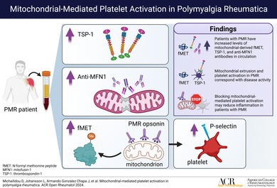
Objective: Platelet activation is thought to participate in polymyalgia rheumatica (PMR) pathogenesis. Upon platelet activation, mitochondria are expelled into the extracellular space. However, whether extracellular mitochondria are present in patients with PMR and whether they can induce platelet activation is not known.
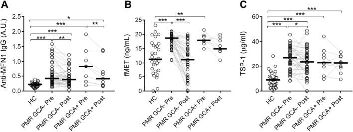
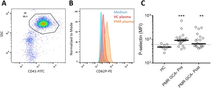
Methods: To investigate this, we measured markers of platelet activation (thrombospondin-1 [TSP-1]), mitochondrial-derived N-formyl methionine peptide (fMET), and autoantibodies directed toward specific mitochondrial antigen mitofusin-1 (MFN1) by enzyme-linked immunosorbent assay in plasma of healthy controls (HCs, n = 30) and patients with PMR without giant cell arteritis (GCA) (n = 45) and patients with PMR with GCA (n = 9) before and after treatment with glucocorticoid therapy. Ultrapure mitochondria were opsonized with plasma from patients with PMR without GCA (n = 45) or HCs (n = 10) and were subsequently incubated with HC platelets. Platelet activation was assessed by P-selectin levels using flow cytometry.
Results: Plasma levels of anti-MFN1 IgG were elevated in patients with PMR with and without GCA before glucocorticoid therapy when compared with HCs (P < 0.01 for both groups). Levels of anti-MFN1 IgG significantly reduced after treatment with glucocorticoids in both groups (P < 0.01). Levels of fMET were also significantly higher in patients with PMR with and without GCA before glucocorticoid therapy in comparison with HCs (P < 0.001 and P < 0.01, respectively). However, the levels of fMET only dropped significantly after therapy in patients with PMR without GCA (P < 0.001). Plasma levels of TSP-1 were elevated in patients with PMR with and without GCA before glucocorticoid therapy when compared to HC (P < 0.001 for both groups). After glucocorticoid therapy, plasma levels of TSP-1 decreased significantly only in patients with PMR without GCA (P = 0.023). Mitochondria opsonized with plasma from patients with PMR without GCA induced higher platelet activation regardless of treatment status as compared with plasma from HCs (P < 0.0001 and P < 0.01 for pretreatment and posttreatment).
Conclusion: Our results indicate increased platelet activation and the presence of mitochondrial antigens and antibodies in the circulation of patients with PMR. Blocking mitochondrial-mediated platelet activation may reduce inflammation in patients with PMR, with potential therapeutic implications.

 求助内容:
求助内容: 应助结果提醒方式:


