{"title":"Vascular supply of the eye: clinical anatomy.","authors":"Omer Karti, Isil Saatci, Ali Osman Saatci","doi":"10.51329/mehdiophthal1509","DOIUrl":null,"url":null,"abstract":"<p><strong>Background: </strong>Eye function is vitally dependent on an adequate blood supply, primarily provided by the ophthalmic artery, an internal carotid artery branch. This review provides an overview of the vascular supply of the eye.</p><p><strong>Methods: </strong>A targeted search of PubMed / MEDLINE was performed using the terms \"central retinal vein,\" \"central retinal artery,\" \"internal carotid artery,\" \"ophthalmic artery,\" \"ophthalmic vein,\" \"posterior ciliary arteries,\" \"retinal capillaries,\" \"vascular supply of the eye,\" \"ocular vascular supply,\" \"external carotid artery,\" and \"vortex vein\". Studies published between 1960 and 2024 were reviewed. Relevant references cited in these publications were also analyzed.</p><p><strong>Results: </strong>Overall, 62 publications were reviewed. The ophthalmic artery branches into several arteries-the central retinal artery supplies the retina, whereas the posterior ciliary arteries supply the posterior choroid and optic nerve. The anterior ciliary arteries mainly supply the conjunctiva, sclera, ciliary body, and iris. Extraocular muscles receive their primary blood supply from the muscular branches of the ophthalmic artery, lacrimal artery, and infraorbital artery. The lacrimal gland is perfused by the lacrimal artery. The eyelids receive blood from both the internal and external carotid arteries. The superficial vascular network of the medial eyelid skin is established primarily through anastomoses between the branches of the internal carotid artery. The superficial vascular network of the lateral upper and lower eyelids is primarily derived from branches emanating from the superficial temporal artery (a branch of the external carotid artery) and the lacrimal artery. Venous drainage follows a complex pathway, beginning with the central retinal vein and the vortex veins, then draining into the ophthalmic veins, and finally into the internal jugular vein.</p><p><strong>Conclusions: </strong>The eye features a complex arterial supply and venous drainage that can vary greatly among individuals. This complex vascular system is critical for the oxygenation and nutrition of ocular tissues and the maintenance of ocular health. The arterial and venous circulation coordinate to support different regions of the eye, including the retina, choroid, and optic nerve. Understanding this intricate vascular network is essential for the diagnosis and treatment of various ocular pathologies. Abnormalities in these pathways can cause substantial problems, including vision loss.</p>","PeriodicalId":36524,"journal":{"name":"Medical Hypothesis, Discovery, and Innovation in Ophthalmology","volume":"13 4","pages":"176-189"},"PeriodicalIF":0.0000,"publicationDate":"2025-02-01","publicationTypes":"Journal Article","fieldsOfStudy":null,"isOpenAccess":false,"openAccessPdf":"https://www.ncbi.nlm.nih.gov/pmc/articles/PMC11890264/pdf/","citationCount":"0","resultStr":null,"platform":"Semanticscholar","paperid":null,"PeriodicalName":"Medical Hypothesis, Discovery, and Innovation in Ophthalmology","FirstCategoryId":"1085","ListUrlMain":"https://doi.org/10.51329/mehdiophthal1509","RegionNum":0,"RegionCategory":null,"ArticlePicture":[],"TitleCN":null,"AbstractTextCN":null,"PMCID":null,"EPubDate":"2024/1/1 0:00:00","PubModel":"eCollection","JCR":"Q2","JCRName":"Medicine","Score":null,"Total":0}
引用次数: 0
Abstract
Background: Eye function is vitally dependent on an adequate blood supply, primarily provided by the ophthalmic artery, an internal carotid artery branch. This review provides an overview of the vascular supply of the eye.
Methods: A targeted search of PubMed / MEDLINE was performed using the terms "central retinal vein," "central retinal artery," "internal carotid artery," "ophthalmic artery," "ophthalmic vein," "posterior ciliary arteries," "retinal capillaries," "vascular supply of the eye," "ocular vascular supply," "external carotid artery," and "vortex vein". Studies published between 1960 and 2024 were reviewed. Relevant references cited in these publications were also analyzed.
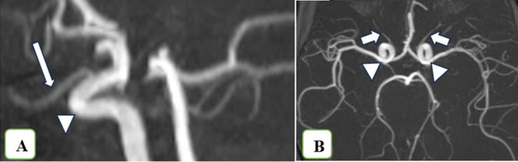
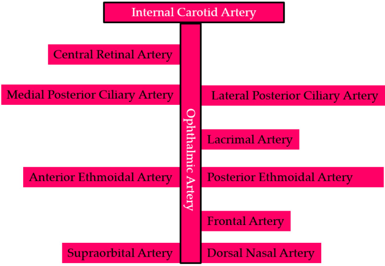
Results: Overall, 62 publications were reviewed. The ophthalmic artery branches into several arteries-the central retinal artery supplies the retina, whereas the posterior ciliary arteries supply the posterior choroid and optic nerve. The anterior ciliary arteries mainly supply the conjunctiva, sclera, ciliary body, and iris. Extraocular muscles receive their primary blood supply from the muscular branches of the ophthalmic artery, lacrimal artery, and infraorbital artery. The lacrimal gland is perfused by the lacrimal artery. The eyelids receive blood from both the internal and external carotid arteries. The superficial vascular network of the medial eyelid skin is established primarily through anastomoses between the branches of the internal carotid artery. The superficial vascular network of the lateral upper and lower eyelids is primarily derived from branches emanating from the superficial temporal artery (a branch of the external carotid artery) and the lacrimal artery. Venous drainage follows a complex pathway, beginning with the central retinal vein and the vortex veins, then draining into the ophthalmic veins, and finally into the internal jugular vein.
Conclusions: The eye features a complex arterial supply and venous drainage that can vary greatly among individuals. This complex vascular system is critical for the oxygenation and nutrition of ocular tissues and the maintenance of ocular health. The arterial and venous circulation coordinate to support different regions of the eye, including the retina, choroid, and optic nerve. Understanding this intricate vascular network is essential for the diagnosis and treatment of various ocular pathologies. Abnormalities in these pathways can cause substantial problems, including vision loss.

 求助内容:
求助内容: 应助结果提醒方式:


