{"title":"Granulomatous liver disease in Thailand: a 20-year retrospective clinicoradiopathological analysis.","authors":"Siwanon Nawalerspanya, Apichat Kaewdech, Naichaya Chamroonkul, Pimsiri Sripongpun","doi":"10.1136/bmjgast-2024-001675","DOIUrl":null,"url":null,"abstract":"<p><strong>Objective: </strong>Granulomatous liver disease (GLD) is a rare condition with various aetiologies and is characterised by the formation of hepatic granulomas. A comprehensive evaluation of GLD from a broad perspective is lacking. We aimed to investigate the aetiology and the clinicoradiopathological characteristics of patients with GLD in recent decades in Thailand.</p><p><strong>Methods: </strong>This retrospective study was conducted at a tertiary care centre in Thailand. All patients who underwent liver biopsy between 2003 and 2023 were reviewed. Patients with a histopathological report of granulomas in liver specimens were included. Clinical presentations, radiological data, and laboratory data closest to the procedure date were also collected.</p><p><strong>Results: </strong>Of the 4384 liver biopsy specimens collected during the study period, 89 (2%) had GLD. Of these, 58.4% were men, with the following aetiologies: 61 (68.5%) infectious, 16 (18%) non-infectious, and 12 (13.5%) undetermined. Common presentations included abnormal liver test results (81.4%) and fever (56.1%). Among infectious granulomas, mycobacterial infections (tuberculosis: 28; non-tuberculous mycobacteria (NTM): 11) were predominant. Compared with other causes, NTM was associated with a significantly lower body mass index, more extragastrointestinal involvement, and lower serum albumin levels. Caseating-type granulomas were also observed in 16% of non-mycobacterial cases. Nearly 40% of patients with GLD demonstrated no focal lesions on liver imaging, whereas multifocal lesions were found in a third of patients.</p><p><strong>Conclusions: </strong>Infectious causes, especially mycobacterial infections, remain the primary aetiology of GLD in Thailand. Granuloma types are not pathognomonic of specific diseases, emphasising the need for extensive evaluation beyond liver biopsy to determine the underlying aetiology.</p>","PeriodicalId":9235,"journal":{"name":"BMJ Open Gastroenterology","volume":"12 1","pages":""},"PeriodicalIF":2.9000,"publicationDate":"2025-03-06","publicationTypes":"Journal Article","fieldsOfStudy":null,"isOpenAccess":false,"openAccessPdf":"https://www.ncbi.nlm.nih.gov/pmc/articles/PMC11887282/pdf/","citationCount":"0","resultStr":null,"platform":"Semanticscholar","paperid":null,"PeriodicalName":"BMJ Open Gastroenterology","FirstCategoryId":"1085","ListUrlMain":"https://doi.org/10.1136/bmjgast-2024-001675","RegionNum":0,"RegionCategory":null,"ArticlePicture":[],"TitleCN":null,"AbstractTextCN":null,"PMCID":null,"EPubDate":"","PubModel":"","JCR":"Q2","JCRName":"GASTROENTEROLOGY & HEPATOLOGY","Score":null,"Total":0}
引用次数: 0
Abstract
Objective: Granulomatous liver disease (GLD) is a rare condition with various aetiologies and is characterised by the formation of hepatic granulomas. A comprehensive evaluation of GLD from a broad perspective is lacking. We aimed to investigate the aetiology and the clinicoradiopathological characteristics of patients with GLD in recent decades in Thailand.
Methods: This retrospective study was conducted at a tertiary care centre in Thailand. All patients who underwent liver biopsy between 2003 and 2023 were reviewed. Patients with a histopathological report of granulomas in liver specimens were included. Clinical presentations, radiological data, and laboratory data closest to the procedure date were also collected.
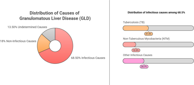
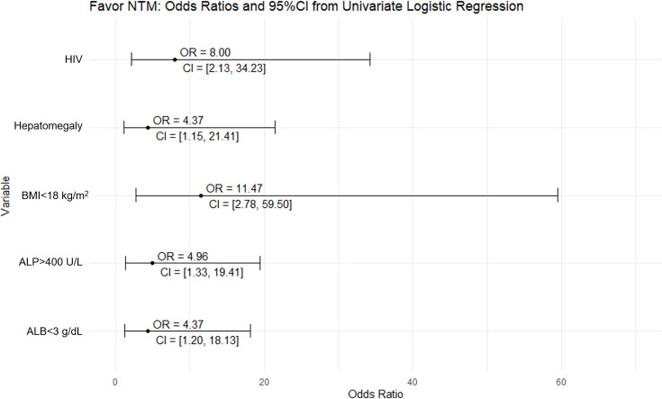
Results: Of the 4384 liver biopsy specimens collected during the study period, 89 (2%) had GLD. Of these, 58.4% were men, with the following aetiologies: 61 (68.5%) infectious, 16 (18%) non-infectious, and 12 (13.5%) undetermined. Common presentations included abnormal liver test results (81.4%) and fever (56.1%). Among infectious granulomas, mycobacterial infections (tuberculosis: 28; non-tuberculous mycobacteria (NTM): 11) were predominant. Compared with other causes, NTM was associated with a significantly lower body mass index, more extragastrointestinal involvement, and lower serum albumin levels. Caseating-type granulomas were also observed in 16% of non-mycobacterial cases. Nearly 40% of patients with GLD demonstrated no focal lesions on liver imaging, whereas multifocal lesions were found in a third of patients.
Conclusions: Infectious causes, especially mycobacterial infections, remain the primary aetiology of GLD in Thailand. Granuloma types are not pathognomonic of specific diseases, emphasising the need for extensive evaluation beyond liver biopsy to determine the underlying aetiology.
期刊介绍:
BMJ Open Gastroenterology is an online-only, peer-reviewed, open access gastroenterology journal, dedicated to publishing high-quality medical research from all disciplines and therapeutic areas of gastroenterology. It is the open access companion journal of Gut and is co-owned by the British Society of Gastroenterology. The journal publishes all research study types, from study protocols to phase I trials to meta-analyses, including small or specialist studies. Publishing procedures are built around continuous publication, publishing research online as soon as the article is ready.

 求助内容:
求助内容: 应助结果提醒方式:


