Mahmoud R Manasra, Rahaf E Farah, Roua E Farah, Sama S Yassin, Shadi A Abuisneina
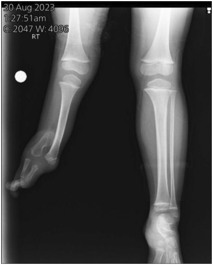
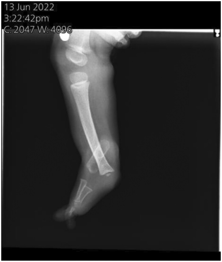
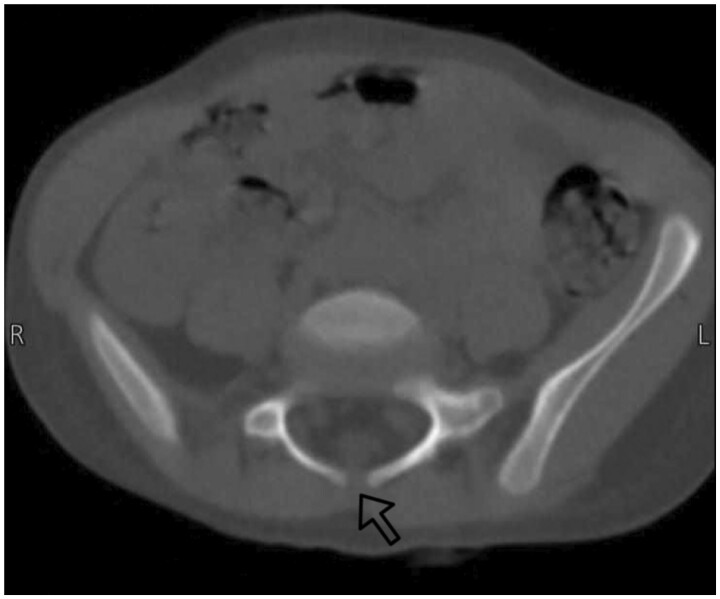
{"title":"A case of congenital fibular hemimelia associated with skeletal and non-skeletal malformations.","authors":"Mahmoud R Manasra, Rahaf E Farah, Roua E Farah, Sama S Yassin, Shadi A Abuisneina","doi":"10.1093/bjrcr/uaaf008","DOIUrl":null,"url":null,"abstract":"<p><p>Fibular hemimelia (FH) is a rare congenital abnormality where the fibula is either totally or partially absent. It can occur alone or alongside other skeletal malformations, and in very few cases, it may occur along with non-skeletal anomalies. A 4-year-old female was diagnosed with unilateral right-sided FH, accompanied by limb shortening, a right-side ankle deformity, valgus foot, and 3 lateral rays that had been totally absent since the first week of birth. And she was incidentally diagnosed with spina bifida occulta at a 3-year-old age. FH is most commonly unilateral and mostly affects the right side, leads to a limb-length discrepancy, and maybe comes as a symptom of a syndrome such as Foetus-Fibula-Ulna syndrome and so on. Risk factors include prenatal history, drugs, and no supplementation intake. Together, these elements could be a contributing factor to our condition. The congenital limb abnormalities may be discovered during pregnancy by sonography. If present, other investigations need to be done to differentiate the diagnosis. Treatment according to degree: mild, moderate, and severe cases. In our case, the type 2 FH characteristic was shown by sonography, accompanied by limb shortening, lateral rays absent, and a non-skeletal anomaly (spina bifida). These anomalies very rarely come with each other at the same time. To the best of our knowledge, this case is exceptional in that FH is present at birth alongside spina bifida.</p>","PeriodicalId":45216,"journal":{"name":"BJR Case Reports","volume":"11 2","pages":"uaaf008"},"PeriodicalIF":0.5000,"publicationDate":"2025-02-18","publicationTypes":"Journal Article","fieldsOfStudy":null,"isOpenAccess":false,"openAccessPdf":"https://www.ncbi.nlm.nih.gov/pmc/articles/PMC11879310/pdf/","citationCount":"0","resultStr":null,"platform":"Semanticscholar","paperid":null,"PeriodicalName":"BJR Case Reports","FirstCategoryId":"1085","ListUrlMain":"https://doi.org/10.1093/bjrcr/uaaf008","RegionNum":0,"RegionCategory":null,"ArticlePicture":[],"TitleCN":null,"AbstractTextCN":null,"PMCID":null,"EPubDate":"2025/3/1 0:00:00","PubModel":"eCollection","JCR":"Q4","JCRName":"RADIOLOGY, NUCLEAR MEDICINE & MEDICAL IMAGING","Score":null,"Total":0}
引用次数: 0
Abstract
Fibular hemimelia (FH) is a rare congenital abnormality where the fibula is either totally or partially absent. It can occur alone or alongside other skeletal malformations, and in very few cases, it may occur along with non-skeletal anomalies. A 4-year-old female was diagnosed with unilateral right-sided FH, accompanied by limb shortening, a right-side ankle deformity, valgus foot, and 3 lateral rays that had been totally absent since the first week of birth. And she was incidentally diagnosed with spina bifida occulta at a 3-year-old age. FH is most commonly unilateral and mostly affects the right side, leads to a limb-length discrepancy, and maybe comes as a symptom of a syndrome such as Foetus-Fibula-Ulna syndrome and so on. Risk factors include prenatal history, drugs, and no supplementation intake. Together, these elements could be a contributing factor to our condition. The congenital limb abnormalities may be discovered during pregnancy by sonography. If present, other investigations need to be done to differentiate the diagnosis. Treatment according to degree: mild, moderate, and severe cases. In our case, the type 2 FH characteristic was shown by sonography, accompanied by limb shortening, lateral rays absent, and a non-skeletal anomaly (spina bifida). These anomalies very rarely come with each other at the same time. To the best of our knowledge, this case is exceptional in that FH is present at birth alongside spina bifida.

 求助内容:
求助内容: 应助结果提醒方式:


