Atlas Haddadi Avval, Suneel Banerjee, John Zielke, Benjamin H Kann, Sabine Mueller, Andreas M Rauschecker
{"title":"Applications of artificial intelligence and advanced imaging in pediatric diffuse midline glioma.","authors":"Atlas Haddadi Avval, Suneel Banerjee, John Zielke, Benjamin H Kann, Sabine Mueller, Andreas M Rauschecker","doi":"10.1093/neuonc/noaf058","DOIUrl":null,"url":null,"abstract":"<p><p>Diffuse midline glioma (DMG) is a rare, aggressive, and fatal tumor that largely occurs in the pediatric population. To improve outcomes, it is important to characterize DMGs, which can be performed via magnetic resonance imaging (MRI) assessment. Recently, artificial intelligence (AI) and advanced imaging have demonstrated their potential to improve the evaluation of various brain tumors, gleaning more information from imaging data than is possible without these methods. This narrative review compiles the existing literature on the intersection of MRI-based AI use and DMG tumors. The applications of AI in DMG revolve around classification and diagnosis, segmentation, radiogenomics, and prognosis/survival prediction. Currently published articles have utilized a wide spectrum of AI algorithms, from traditional machine learning and radiomics to neural networks. Challenges include the lack of cohorts of DMG patients with publicly available, multi-institutional, multimodal imaging and genomics datasets as well as the overall rarity of the disease. As an adjunct to AI, advanced MRI techniques, including diffusion-weighted imaging, perfusion-weighted imaging, and Magnetic Resonance Spectroscopy (MRS), as well as positron emission tomography (PET), provide additional insights into DMGs. Establishing AI models in conjunction with advanced imaging modalities has the potential to push clinical practice toward precision medicine.</p>","PeriodicalId":19377,"journal":{"name":"Neuro-oncology","volume":" ","pages":"1419-1433"},"PeriodicalIF":13.4000,"publicationDate":"2025-07-30","publicationTypes":"Journal Article","fieldsOfStudy":null,"isOpenAccess":false,"openAccessPdf":"https://www.ncbi.nlm.nih.gov/pmc/articles/PMC12309720/pdf/","citationCount":"0","resultStr":null,"platform":"Semanticscholar","paperid":null,"PeriodicalName":"Neuro-oncology","FirstCategoryId":"3","ListUrlMain":"https://doi.org/10.1093/neuonc/noaf058","RegionNum":1,"RegionCategory":"医学","ArticlePicture":[],"TitleCN":null,"AbstractTextCN":null,"PMCID":null,"EPubDate":"","PubModel":"","JCR":"Q1","JCRName":"CLINICAL NEUROLOGY","Score":null,"Total":0}
引用次数: 0
Abstract
Diffuse midline glioma (DMG) is a rare, aggressive, and fatal tumor that largely occurs in the pediatric population. To improve outcomes, it is important to characterize DMGs, which can be performed via magnetic resonance imaging (MRI) assessment. Recently, artificial intelligence (AI) and advanced imaging have demonstrated their potential to improve the evaluation of various brain tumors, gleaning more information from imaging data than is possible without these methods. This narrative review compiles the existing literature on the intersection of MRI-based AI use and DMG tumors. The applications of AI in DMG revolve around classification and diagnosis, segmentation, radiogenomics, and prognosis/survival prediction. Currently published articles have utilized a wide spectrum of AI algorithms, from traditional machine learning and radiomics to neural networks. Challenges include the lack of cohorts of DMG patients with publicly available, multi-institutional, multimodal imaging and genomics datasets as well as the overall rarity of the disease. As an adjunct to AI, advanced MRI techniques, including diffusion-weighted imaging, perfusion-weighted imaging, and Magnetic Resonance Spectroscopy (MRS), as well as positron emission tomography (PET), provide additional insights into DMGs. Establishing AI models in conjunction with advanced imaging modalities has the potential to push clinical practice toward precision medicine.
期刊介绍:
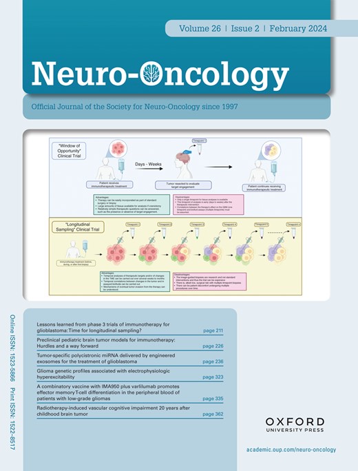
Neuro-Oncology, the official journal of the Society for Neuro-Oncology, has been published monthly since January 2010. Affiliated with the Japan Society for Neuro-Oncology and the European Association of Neuro-Oncology, it is a global leader in the field.
The journal is committed to swiftly disseminating high-quality information across all areas of neuro-oncology. It features peer-reviewed articles, reviews, symposia on various topics, abstracts from annual meetings, and updates from neuro-oncology societies worldwide.
 求助内容:
求助内容: 应助结果提醒方式:


