{"title":"Tibial morphological difference between metal augmentation and actual tibia for revision total knee arthroplasty.","authors":"Yushi Sakamoto, Shinichiro Nakamura, Yugo Morita, Shinichi Kuriyama, Kohei Nishitani, Sayako Sakai, Yuki Shinya, Shuichi Matsuda","doi":"10.1186/s43019-025-00262-9","DOIUrl":null,"url":null,"abstract":"<p><strong>Background: </strong>An overhang of the tibial component can cause irritation of the surrounding soft tissues, whereas an underhang is associated with risks of tibial bone resorption. It is not well known whether the tapering angle of currently available blocks at medial, lateral, anterior, and posterior sides matches the actual shape of the proximal tibia. The purpose of this study was to analyze the bony contour of the proximal tibia and measure the tapering angle to examine whether the angle of currently available metal augmentation blocks matches the actual tibia.</p><p><strong>Methods: </strong>Computed tomography of the lower extremities was performed on 100 consecutive knees, and three-dimensional images of the tibia were reconstructed. The primary resection level was determined on the basis of a plane 10 mm below the center of the lateral tibial plateau. The assumed levels were set to 5, 10, 15, and 20 mm below the primary resection level. All points that were 5, 10, 15, or 20 mm below were projected onto the primary resection surface, and the reduction value from the primary level to each level was measured. The tapering angle was calculated on the basis of the reduction value from the primary level to each resection surface at eight areas and compared with the angle of currently available metal augmentation acquired from the company. The distances of mismatch between the metal augmentation and the bone surface were calculated.</p><p><strong>Results: </strong>The tapering angle on the medial and lateral sides increased with the more distal resection level, which was up to 30° at the 20 mm level. The tapering angle on the posterior side also increased with the more distal resection level, which was approximately 40° at the 20 mm level. The tapering angle of the current implant was smaller than that of the original tibial morphology. The distances of mismatch varied between implants in which the maximum distance was up to 11.3 mm in the 15 mm augmentation.</p><p><strong>Conclusions: </strong>The design of current metal augmentation differs from the morphology of the proximal tibia. Surgeons should pay attention to the size mismatch between the femoral and tibial components during revision total knee arthroplasty (TKA).</p>","PeriodicalId":36317,"journal":{"name":"Knee Surgery and Related Research","volume":"37 1","pages":"10"},"PeriodicalIF":4.4000,"publicationDate":"2025-03-03","publicationTypes":"Journal Article","fieldsOfStudy":null,"isOpenAccess":false,"openAccessPdf":"https://www.ncbi.nlm.nih.gov/pmc/articles/PMC11874112/pdf/","citationCount":"0","resultStr":null,"platform":"Semanticscholar","paperid":null,"PeriodicalName":"Knee Surgery and Related Research","FirstCategoryId":"1085","ListUrlMain":"https://doi.org/10.1186/s43019-025-00262-9","RegionNum":0,"RegionCategory":null,"ArticlePicture":[],"TitleCN":null,"AbstractTextCN":null,"PMCID":null,"EPubDate":"","PubModel":"","JCR":"Q2","JCRName":"Medicine","Score":null,"Total":0}
引用次数: 0
Abstract
Background: An overhang of the tibial component can cause irritation of the surrounding soft tissues, whereas an underhang is associated with risks of tibial bone resorption. It is not well known whether the tapering angle of currently available blocks at medial, lateral, anterior, and posterior sides matches the actual shape of the proximal tibia. The purpose of this study was to analyze the bony contour of the proximal tibia and measure the tapering angle to examine whether the angle of currently available metal augmentation blocks matches the actual tibia.
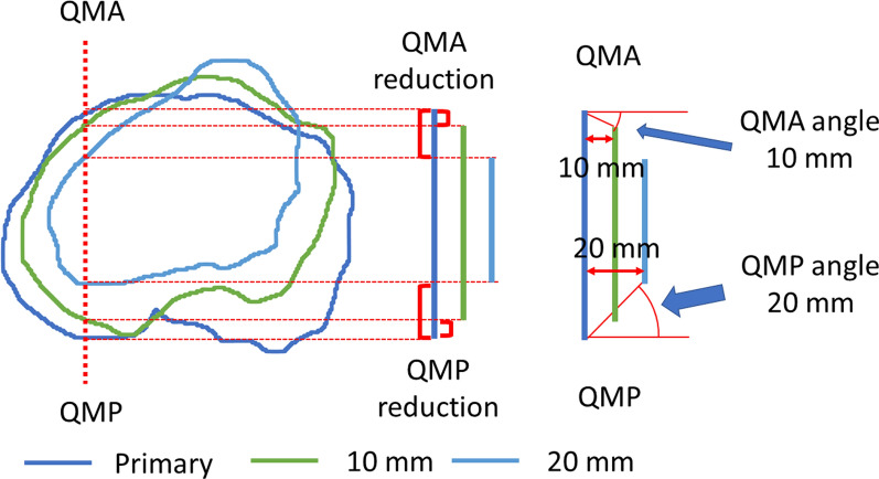
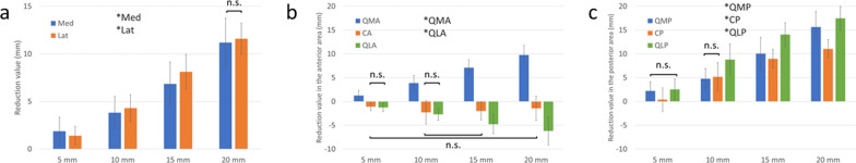
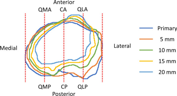
Methods: Computed tomography of the lower extremities was performed on 100 consecutive knees, and three-dimensional images of the tibia were reconstructed. The primary resection level was determined on the basis of a plane 10 mm below the center of the lateral tibial plateau. The assumed levels were set to 5, 10, 15, and 20 mm below the primary resection level. All points that were 5, 10, 15, or 20 mm below were projected onto the primary resection surface, and the reduction value from the primary level to each level was measured. The tapering angle was calculated on the basis of the reduction value from the primary level to each resection surface at eight areas and compared with the angle of currently available metal augmentation acquired from the company. The distances of mismatch between the metal augmentation and the bone surface were calculated.
Results: The tapering angle on the medial and lateral sides increased with the more distal resection level, which was up to 30° at the 20 mm level. The tapering angle on the posterior side also increased with the more distal resection level, which was approximately 40° at the 20 mm level. The tapering angle of the current implant was smaller than that of the original tibial morphology. The distances of mismatch varied between implants in which the maximum distance was up to 11.3 mm in the 15 mm augmentation.
Conclusions: The design of current metal augmentation differs from the morphology of the proximal tibia. Surgeons should pay attention to the size mismatch between the femoral and tibial components during revision total knee arthroplasty (TKA).

 求助内容:
求助内容: 应助结果提醒方式:


