Martin B Schilder, Stefano Mandija, Sarah M Jacobs, Jordi P D Kleinloog, Hanna Liu, Oscar van der Heide, Beyza Köktaş, Federico D'Agata, Vera C W Keil, Evert-Jan P A Vonken, Jan Willem Dankbaar, Jeroen Hendrikse, Tom J Snijders, Cornelis A T van den Berg, Anja G van der Kolk, Alessandro Sbrizzi
{"title":"Fast whole brain relaxometry with Magnetic Resonance Spin TomogrAphy in Time-domain (MR-STAT) at 3 T: a retrospective cohort study.","authors":"Martin B Schilder, Stefano Mandija, Sarah M Jacobs, Jordi P D Kleinloog, Hanna Liu, Oscar van der Heide, Beyza Köktaş, Federico D'Agata, Vera C W Keil, Evert-Jan P A Vonken, Jan Willem Dankbaar, Jeroen Hendrikse, Tom J Snijders, Cornelis A T van den Berg, Anja G van der Kolk, Alessandro Sbrizzi","doi":"10.1007/s10334-025-01237-3","DOIUrl":null,"url":null,"abstract":"<p><strong>Objective: </strong>To report T<sub>1</sub>/T<sub>2</sub>-values of normal and normal appearing brain tissues (NBTs, healthy volunteers; NABTs, patients) acquired with a whole-brain 5-minute Magnetic Resonance Spin TomogrAphy in Time-domain (MR-STAT) protocol, and to explore relaxometry behavior in a brain tumor and in a multiple sclerosis patient.</p><p><strong>Methods: </strong>MR-STAT was acquired in 49 participants (39 patients with neurological pathologies, age range: 21-79 years) at 3 T. Mean T<sub>1</sub>/T<sub>2</sub>-values were computed for: normal and normal appearing grey matter (NGM/NAGM)/white matter (NWM/NAWM)/thalamus/putamen/caudate nucleus (CN)/globus pallidus (GP). Differences between sex, brain lobes, and left/right were assessed. The age-dependency of T<sub>1</sub>/T<sub>2</sub>-values in N(A)BTs was investigated. Relaxometry analysis was performed in two clinical case examples.</p><p><strong>Results: </strong>Mean (standard deviation) T<sub>1</sub>/T<sub>2</sub>-values were measured in N(A)GM = 1086(73)/74(9) ms; N(A)WM = 658(24)/48(3) ms; thalamus = 783(51)/42(4) ms; putamen = 863(40)/46(3) ms; CN = 1042(97)/63(9) ms; GP = 652(36)/36(3) ms. Differences between sex were not significant. T<sub>1</sub>/T<sub>2</sub>-values between the left/right parietal lobe and the left/right temporal lobe were significantly different. The quadratic age-dependency of T<sub>1</sub>-values in the CN (p = 0.00039) and GP (p = 0.00037), and of T<sub>2</sub>-values in the thalamus (p = 0.00044) and GP (p = 0.003) were significant. Pathological tissues could be discerned from NABTs using T<sub>1</sub>/T<sub>2</sub>-values.</p><p><strong>Discussion: </strong>T<sub>1</sub>/T<sub>2</sub>-values and data trends agree with literature, supporting the validity of MR-STAT as a clinical option for fast relaxometry despite the relatively low number of subjects in the study. Future work should aim to include healthy participants of a wider age-range and to include B<sub>1</sub>-field corrections.</p>","PeriodicalId":18067,"journal":{"name":"Magnetic Resonance Materials in Physics, Biology and Medicine","volume":" ","pages":"333-345"},"PeriodicalIF":2.5000,"publicationDate":"2025-04-01","publicationTypes":"Journal Article","fieldsOfStudy":null,"isOpenAccess":false,"openAccessPdf":"https://www.ncbi.nlm.nih.gov/pmc/articles/PMC11914305/pdf/","citationCount":"0","resultStr":null,"platform":"Semanticscholar","paperid":null,"PeriodicalName":"Magnetic Resonance Materials in Physics, Biology and Medicine","FirstCategoryId":"3","ListUrlMain":"https://doi.org/10.1007/s10334-025-01237-3","RegionNum":4,"RegionCategory":"医学","ArticlePicture":[],"TitleCN":null,"AbstractTextCN":null,"PMCID":null,"EPubDate":"2025/3/4 0:00:00","PubModel":"Epub","JCR":"Q3","JCRName":"RADIOLOGY, NUCLEAR MEDICINE & MEDICAL IMAGING","Score":null,"Total":0}
引用次数: 0
Abstract
Objective: To report T1/T2-values of normal and normal appearing brain tissues (NBTs, healthy volunteers; NABTs, patients) acquired with a whole-brain 5-minute Magnetic Resonance Spin TomogrAphy in Time-domain (MR-STAT) protocol, and to explore relaxometry behavior in a brain tumor and in a multiple sclerosis patient.
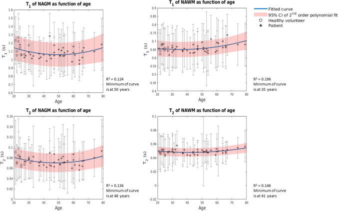
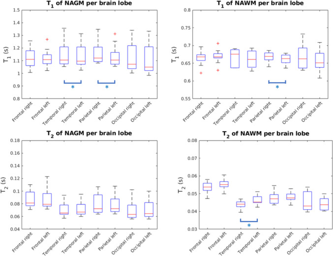
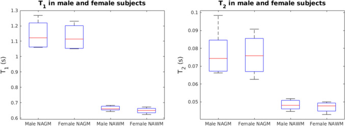
Methods: MR-STAT was acquired in 49 participants (39 patients with neurological pathologies, age range: 21-79 years) at 3 T. Mean T1/T2-values were computed for: normal and normal appearing grey matter (NGM/NAGM)/white matter (NWM/NAWM)/thalamus/putamen/caudate nucleus (CN)/globus pallidus (GP). Differences between sex, brain lobes, and left/right were assessed. The age-dependency of T1/T2-values in N(A)BTs was investigated. Relaxometry analysis was performed in two clinical case examples.
Results: Mean (standard deviation) T1/T2-values were measured in N(A)GM = 1086(73)/74(9) ms; N(A)WM = 658(24)/48(3) ms; thalamus = 783(51)/42(4) ms; putamen = 863(40)/46(3) ms; CN = 1042(97)/63(9) ms; GP = 652(36)/36(3) ms. Differences between sex were not significant. T1/T2-values between the left/right parietal lobe and the left/right temporal lobe were significantly different. The quadratic age-dependency of T1-values in the CN (p = 0.00039) and GP (p = 0.00037), and of T2-values in the thalamus (p = 0.00044) and GP (p = 0.003) were significant. Pathological tissues could be discerned from NABTs using T1/T2-values.
Discussion: T1/T2-values and data trends agree with literature, supporting the validity of MR-STAT as a clinical option for fast relaxometry despite the relatively low number of subjects in the study. Future work should aim to include healthy participants of a wider age-range and to include B1-field corrections.
期刊介绍:
MAGMA is a multidisciplinary international journal devoted to the publication of articles on all aspects of magnetic resonance techniques and their applications in medicine and biology. MAGMA currently publishes research papers, reviews, letters to the editor, and commentaries, six times a year. The subject areas covered by MAGMA include:
advances in materials, hardware and software in magnetic resonance technology,
new developments and results in research and practical applications of magnetic resonance imaging and spectroscopy related to biology and medicine,
study of animal models and intact cells using magnetic resonance,
reports of clinical trials on humans and clinical validation of magnetic resonance protocols.

 求助内容:
求助内容: 应助结果提醒方式:


