Bone density comparison between the normal pedicle trajectory, cortical bone trajectory, and modified cortical bone trajectory using computed tomography: a cross-sectional study.
{"title":"Bone density comparison between the normal pedicle trajectory, cortical bone trajectory, and modified cortical bone trajectory using computed tomography: a cross-sectional study.","authors":"Bikash Parajuli, Dipak Shrestha, Kuniyoshi Abumi, Sabik Kayastha, Jagadish Thapa, Sanjay Sharma, Suman Lamichhane, Christian Deininger","doi":"10.31616/asj.2024.0377","DOIUrl":null,"url":null,"abstract":"<p><strong>Study design: </strong>Cross-sectional study.</p><p><strong>Purpose: </strong>To compare bone density by computed tomography Hounsfield unit (CTHU) between the original pedicle trajectory (OPT), cortical bone trajectory (CBT), and modified cortical bone trajectory (MCBT).</p><p><strong>Overview of literature: </strong>The significant pullout strength in CBT is believed to be due to increased screw-cortical bone contact; however, it allows for shorter/less-diameter screw placement, and the fixation is limited to the posterior one-third of the vertebral body, compromising the screw anchorage in the anterior vertebra.</p><p><strong>Methods: </strong>Computed tomography transverse sections of the L1-L5 (1,000 vertebrae) of 200 patients were cut into three planes: (1) horizontal to the pedicle, representing the plane for OPT; (2) in the caudocranial plane in the sagittal plane and divergent in the transverse plane representing the CBT; and (3) the caudocranial plane in the sagittal plane and parallel in the transverse plane representing the MCBT. For each trajectory, the CTHU of four points, namely, posterior cortex, mid-pedicle, midbody, and anterior cortex, were compared within the area of screw insertion.</p><p><strong>Results: </strong>The mean CTHUs of OPT, CBT, and MCBT were 354.2±70 HU, 529.9±75 HU, and 457.3±90 HU, respectively (p <0.01). The CTHU of the posterior cortex in MCBT was 65.6% higher than that in OPT and 14.9% lower than that in CBT. A comparable decline in CTHU with age was noted in CBT and MCBT (Pearson's r : -0.20 vs. -0.22; adjusted R 2: 0.040 vs. 0.047). However, a higher decline in CTHU with age was observed in OPT (Pearson's r =-0.38, adjusted R 2=0.14).</p><p><strong>Conclusions: </strong>MCBT has a significantly higher CTHU than OPT. The density in the posterior cortex in MCBT is comparable to that in the CBT trajectory. MCBT appears to be an alternative trajectory for lumbar spine fixation.</p>","PeriodicalId":8555,"journal":{"name":"Asian Spine Journal","volume":" ","pages":"372-379"},"PeriodicalIF":2.7000,"publicationDate":"2025-06-01","publicationTypes":"Journal Article","fieldsOfStudy":null,"isOpenAccess":false,"openAccessPdf":"https://www.ncbi.nlm.nih.gov/pmc/articles/PMC12242248/pdf/","citationCount":"0","resultStr":null,"platform":"Semanticscholar","paperid":null,"PeriodicalName":"Asian Spine Journal","FirstCategoryId":"1085","ListUrlMain":"https://doi.org/10.31616/asj.2024.0377","RegionNum":0,"RegionCategory":null,"ArticlePicture":[],"TitleCN":null,"AbstractTextCN":null,"PMCID":null,"EPubDate":"2025/3/4 0:00:00","PubModel":"Epub","JCR":"Q2","JCRName":"ORTHOPEDICS","Score":null,"Total":0}
引用次数: 0
Abstract
Study design: Cross-sectional study.
Purpose: To compare bone density by computed tomography Hounsfield unit (CTHU) between the original pedicle trajectory (OPT), cortical bone trajectory (CBT), and modified cortical bone trajectory (MCBT).
Overview of literature: The significant pullout strength in CBT is believed to be due to increased screw-cortical bone contact; however, it allows for shorter/less-diameter screw placement, and the fixation is limited to the posterior one-third of the vertebral body, compromising the screw anchorage in the anterior vertebra.
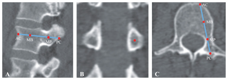
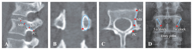
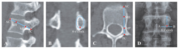
Methods: Computed tomography transverse sections of the L1-L5 (1,000 vertebrae) of 200 patients were cut into three planes: (1) horizontal to the pedicle, representing the plane for OPT; (2) in the caudocranial plane in the sagittal plane and divergent in the transverse plane representing the CBT; and (3) the caudocranial plane in the sagittal plane and parallel in the transverse plane representing the MCBT. For each trajectory, the CTHU of four points, namely, posterior cortex, mid-pedicle, midbody, and anterior cortex, were compared within the area of screw insertion.
Results: The mean CTHUs of OPT, CBT, and MCBT were 354.2±70 HU, 529.9±75 HU, and 457.3±90 HU, respectively (p <0.01). The CTHU of the posterior cortex in MCBT was 65.6% higher than that in OPT and 14.9% lower than that in CBT. A comparable decline in CTHU with age was noted in CBT and MCBT (Pearson's r : -0.20 vs. -0.22; adjusted R 2: 0.040 vs. 0.047). However, a higher decline in CTHU with age was observed in OPT (Pearson's r =-0.38, adjusted R 2=0.14).
Conclusions: MCBT has a significantly higher CTHU than OPT. The density in the posterior cortex in MCBT is comparable to that in the CBT trajectory. MCBT appears to be an alternative trajectory for lumbar spine fixation.

 求助内容:
求助内容: 应助结果提醒方式:


