{"title":"A Rare Intervention in a Rare Disease: Simultaneous Bilateral Keratoplasty in Bilateral <i>Acanthamoeba</i> Keratitis.","authors":"İlayda Korkmaz, Nihat Furkan Eratılgan, Cem Şimşek, Banu Yaman, Sait Eğrilmez, Özlem Barut Selver","doi":"10.4274/tjo.galenos.2024.23934","DOIUrl":null,"url":null,"abstract":"<p><p>The purpose of this report is to present simultaneous bilateral penetrating keratoplasty (PK) in <i>Acanthamoeba</i> keratitis (AK). A 42-year-old male with keratoconus, wearing bilateral hybrid contact lenses, presented with pain in the left eye. He had a history of intrastromal corneal ring segment placement in the right and PK in the left eye. His best corrected visual acuity (BCVA) was 20/640 in the right eye and 20/2000 in the left. Slit-lamp examination revealed a ring-shaped infiltration on the left. Despite two months of broad-spectrum topical antibiotic therapy, microbiological examination of corneal scraping samples was repeated but revealed no evidence of microbial agents. <i>In vivo</i> confocal microscopy findings were not compatible with AK. During the follow-up, corneal infiltration and stromal melt were observed in the right eye. Corneal scraping samples from the right eye were sent for microbiological examination, but again no microbial agents were identified. Histopathological examination revealed spherical cysts consistent with AK. Corneal perforation developed in the right eye, while simultaneous wound dehiscence occurred in the left eye. Since the patient had a history of renal failure, simultaneous bilateral tectonic-therapeutic PK was performed to minimize the risks arising from general anesthesia. Postoperative BCVA was 20/50 in the right eye and 20/125 in the left eye at 6 months. Diagnostic tools can be misleading in eyes with altered anatomy. Careful examination and a timely decision to perform tectonic-therapeutic PK are vital in preventing devastating complications.</p>","PeriodicalId":23373,"journal":{"name":"Turkish Journal of Ophthalmology","volume":"55 1","pages":"49-52"},"PeriodicalIF":0.0000,"publicationDate":"2025-02-27","publicationTypes":"Journal Article","fieldsOfStudy":null,"isOpenAccess":false,"openAccessPdf":"https://www.ncbi.nlm.nih.gov/pmc/articles/PMC11866989/pdf/","citationCount":"0","resultStr":null,"platform":"Semanticscholar","paperid":null,"PeriodicalName":"Turkish Journal of Ophthalmology","FirstCategoryId":"1085","ListUrlMain":"https://doi.org/10.4274/tjo.galenos.2024.23934","RegionNum":0,"RegionCategory":null,"ArticlePicture":[],"TitleCN":null,"AbstractTextCN":null,"PMCID":null,"EPubDate":"","PubModel":"","JCR":"Q3","JCRName":"Medicine","Score":null,"Total":0}
引用次数: 0
Abstract
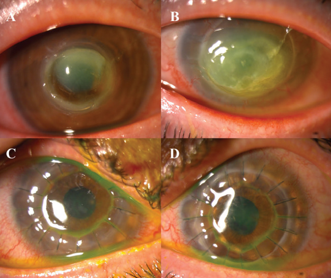
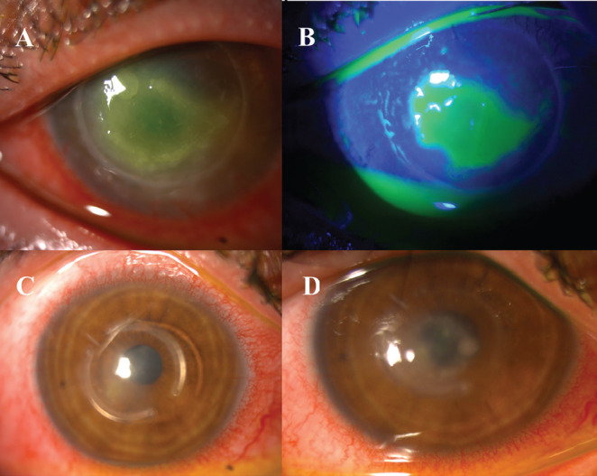
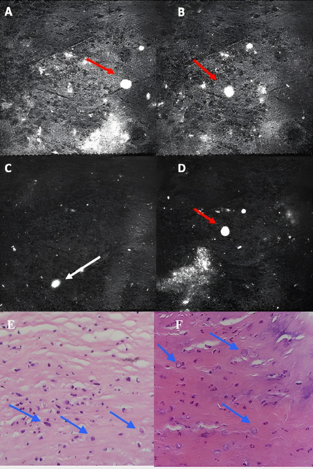
The purpose of this report is to present simultaneous bilateral penetrating keratoplasty (PK) in Acanthamoeba keratitis (AK). A 42-year-old male with keratoconus, wearing bilateral hybrid contact lenses, presented with pain in the left eye. He had a history of intrastromal corneal ring segment placement in the right and PK in the left eye. His best corrected visual acuity (BCVA) was 20/640 in the right eye and 20/2000 in the left. Slit-lamp examination revealed a ring-shaped infiltration on the left. Despite two months of broad-spectrum topical antibiotic therapy, microbiological examination of corneal scraping samples was repeated but revealed no evidence of microbial agents. In vivo confocal microscopy findings were not compatible with AK. During the follow-up, corneal infiltration and stromal melt were observed in the right eye. Corneal scraping samples from the right eye were sent for microbiological examination, but again no microbial agents were identified. Histopathological examination revealed spherical cysts consistent with AK. Corneal perforation developed in the right eye, while simultaneous wound dehiscence occurred in the left eye. Since the patient had a history of renal failure, simultaneous bilateral tectonic-therapeutic PK was performed to minimize the risks arising from general anesthesia. Postoperative BCVA was 20/50 in the right eye and 20/125 in the left eye at 6 months. Diagnostic tools can be misleading in eyes with altered anatomy. Careful examination and a timely decision to perform tectonic-therapeutic PK are vital in preventing devastating complications.
期刊介绍:
The Turkish Journal of Ophthalmology (TJO) is the only scientific periodical publication of the Turkish Ophthalmological Association and has been published since January 1929. In its early years, the journal was published in Turkish and French. Although there were temporary interruptions in the publication of the journal due to various challenges, the Turkish Journal of Ophthalmology has been published continually from 1971 to the present. The target audience includes specialists and physicians in training in ophthalmology in all relevant disciplines.

 求助内容:
求助内容: 应助结果提醒方式:


