Mariwan F Abdalfatah, Abdullah A Shareef, Lana S Saleh, Mustafa F Rajab, Shukur W Smail, Saman S Abdulla, Harem Khdir Awla, Rivan H Ishaac, Kazhal S Ibrahim, Mazyar J Ahmed, Fairuz A Kakasur, Khder Hussein Rasul
{"title":"The Association of <i>VDR/FokI</i> Gene Polymorphism and Protein Expression With Histopathological Alterations in Patients With Thyroid Colloid Nodule.","authors":"Mariwan F Abdalfatah, Abdullah A Shareef, Lana S Saleh, Mustafa F Rajab, Shukur W Smail, Saman S Abdulla, Harem Khdir Awla, Rivan H Ishaac, Kazhal S Ibrahim, Mazyar J Ahmed, Fairuz A Kakasur, Khder Hussein Rasul","doi":"10.1155/ancp/6796922","DOIUrl":null,"url":null,"abstract":"<p><p><b>Objective:</b> Colloid nodules are common and benign thyroid lesions that usually progress slowly and are asymptomatic. It requires follow-up because untreated colloid nodules may develop into malignant tumor. The study aimed to examine the contributions of vitamin D receptor (VDR) expression, VDR/FokI (rs2228570) genotypes, and serum vitamin D level to the susceptibility to colloid nodules. <b>Methods:</b> One hundred forty subjects (80 patients and 60 controls) were enrolled and VDR FokI was determined by PCR in formalin fixed paraffin embedded (FFPE) blocks of the patients and blood of controls. Moreover, VDR protein expression was evaluated by immunohistochemistry using specific VDR monoclonal antibody in the tissue sections of patients and serum vitamin D were measured simultaneously using enzyme-linked immunosorbent assay (ELISA). <b>Results:</b> Sixty-two (77.5%) cases showed strong immunoreactivity score (IRS) of cytoplasmic staining. Strong IRS were significantly observed in samples with larger nodule size (<i>p</i> value: 0.0094), multinodules (<i>p</i> value: 0.0054), and carriers of CC genotypes (<i>p</i> value: 0.0034). TT homozygous genotype revealed significantly (<i>p</i> value: 0.029 and odds ratio (OR): 0.11) protective factor for colloid nodules. In addition, nodule size was significantly (<i>p</i> value: 0.016) larger among CC carriers. Moreover, vitamin D level and category were nonsignificantly difference between patients and controls. <b>Conclusion:</b> Our results reveal prominent cytoplasmic VDR expression, suggesting a distinct distribution pattern and offering valuable insights into its potential role in colloid nodules. VDR expression increases with increasing size and number of nodules. Regarding FokI genotypes, TT genotype was less likely to develop colloid nodule. These findings contribute to our understanding of cellular characteristics of this condition and may have implications for future research and clinical management.</p>","PeriodicalId":49326,"journal":{"name":"Analytical Cellular Pathology","volume":"2025 ","pages":"6796922"},"PeriodicalIF":2.7000,"publicationDate":"2025-02-18","publicationTypes":"Journal Article","fieldsOfStudy":null,"isOpenAccess":false,"openAccessPdf":"https://www.ncbi.nlm.nih.gov/pmc/articles/PMC11858711/pdf/","citationCount":"0","resultStr":null,"platform":"Semanticscholar","paperid":null,"PeriodicalName":"Analytical Cellular Pathology","FirstCategoryId":"3","ListUrlMain":"https://doi.org/10.1155/ancp/6796922","RegionNum":4,"RegionCategory":"医学","ArticlePicture":[],"TitleCN":null,"AbstractTextCN":null,"PMCID":null,"EPubDate":"2025/1/1 0:00:00","PubModel":"eCollection","JCR":"Q3","JCRName":"CELL BIOLOGY","Score":null,"Total":0}
引用次数: 0
Abstract
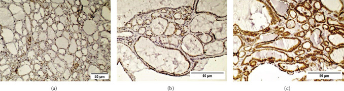
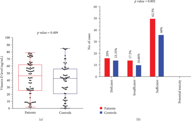
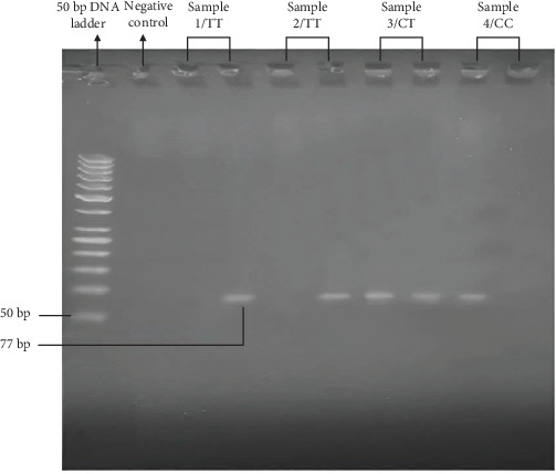
Objective: Colloid nodules are common and benign thyroid lesions that usually progress slowly and are asymptomatic. It requires follow-up because untreated colloid nodules may develop into malignant tumor. The study aimed to examine the contributions of vitamin D receptor (VDR) expression, VDR/FokI (rs2228570) genotypes, and serum vitamin D level to the susceptibility to colloid nodules. Methods: One hundred forty subjects (80 patients and 60 controls) were enrolled and VDR FokI was determined by PCR in formalin fixed paraffin embedded (FFPE) blocks of the patients and blood of controls. Moreover, VDR protein expression was evaluated by immunohistochemistry using specific VDR monoclonal antibody in the tissue sections of patients and serum vitamin D were measured simultaneously using enzyme-linked immunosorbent assay (ELISA). Results: Sixty-two (77.5%) cases showed strong immunoreactivity score (IRS) of cytoplasmic staining. Strong IRS were significantly observed in samples with larger nodule size (p value: 0.0094), multinodules (p value: 0.0054), and carriers of CC genotypes (p value: 0.0034). TT homozygous genotype revealed significantly (p value: 0.029 and odds ratio (OR): 0.11) protective factor for colloid nodules. In addition, nodule size was significantly (p value: 0.016) larger among CC carriers. Moreover, vitamin D level and category were nonsignificantly difference between patients and controls. Conclusion: Our results reveal prominent cytoplasmic VDR expression, suggesting a distinct distribution pattern and offering valuable insights into its potential role in colloid nodules. VDR expression increases with increasing size and number of nodules. Regarding FokI genotypes, TT genotype was less likely to develop colloid nodule. These findings contribute to our understanding of cellular characteristics of this condition and may have implications for future research and clinical management.
期刊介绍:
Analytical Cellular Pathology is a peer-reviewed, Open Access journal that provides a forum for scientists, medical practitioners and pathologists working in the area of cellular pathology. The journal publishes original research articles, review articles, and clinical studies related to cytology, carcinogenesis, cell receptors, biomarkers, diagnostic pathology, immunopathology, and hematology.
 求助内容:
求助内容: 应助结果提醒方式:


