Laura Khatib, Julie Pique, Nicolas Lundahl Ciano-Petersen, Guillaume Criton, Cristina Birzu, Mélodie Aubart, Marie Benaiteau, Geraldine Picard, Romain Marignier, Clarisse Carra-Dalliere, Xavier Ayrignac, Dimitri Psimaras, Pierre M Labauge, Jerome Honnorat, Francois Cotton, Bastien Joubert
{"title":"Abnormal Brain MRI in Anti-NMDA Receptor Encephalitis: Clinical and Prognostic Implications.","authors":"Laura Khatib, Julie Pique, Nicolas Lundahl Ciano-Petersen, Guillaume Criton, Cristina Birzu, Mélodie Aubart, Marie Benaiteau, Geraldine Picard, Romain Marignier, Clarisse Carra-Dalliere, Xavier Ayrignac, Dimitri Psimaras, Pierre M Labauge, Jerome Honnorat, Francois Cotton, Bastien Joubert","doi":"10.1212/NXI.0000000000200378","DOIUrl":null,"url":null,"abstract":"<p><strong>Background and objectives: </strong>Abnormal brain MRI is associated with poor outcomes in anti-N-methyl-d-aspartate receptor encephalitis (NMDARE). We aimed to characterize the lesions on brain MRI in NMDARE and to assess the clinical and prognostic associations.</p><p><strong>Methods: </strong>This retrospective cohort study included patients with NMDARE identified at the French Reference Center for Autoimmune Encephalitis, with at least a one-year follow-up, and with available brain MRI results. In case of brain extralimbic lesion, the image files were reviewed when available. Clinical data were collected from medical records. Multivariable logistic regression analysis was used to study the outcomes at 2-year follow-up; recovery was defined as modified Rankin Scale score ≤1.</p><p><strong>Results: </strong>Among the 255 patients included, 37 (14.5%) had limbic hyperintensities and 41 (16.1%) had extralimbic lesions that included multiple sclerosis (MS)-like lesions (14/41, 34.1%); extensive lesions (5/41, 12.2%); and poorly demarcated fluffy lesions, either multifocal (10/41, 24.4%) or involving the cerebral cortex or cerebellum (6/41 each, 14.6%). Extralimbic lesions coexisting with limbic lesions (19/41 patients, 46.3%) were mostly fluffy lesions (11/19, 57.9%). Ten patients had overlapping demyelinating syndromes: 4 with MS, 4 with myelin oligodendrocyte glycoprotein-associated disorder, and 2 with neuromyelitis optica spectrum disorder; all had MS-like (7/10 patients) or extensive (3/10 patients) lesions, and none had fluffy lesions. Extralimbic lesions were associated with symptoms nontypical for NMDARE (23/41, 56.1%, <i>p</i> < 0.001), especially cerebellar ataxia (17/41, 41.5%) and motor impairment (12/41, 29.3%). At 2 years, patients with MS-like or extensive lesions had a lower recovery rate (5/12, 41.7%, and 1/4, 25%, respectively) compared with the patients without extralimbic lesions (124/162, 76.5%; <i>p</i> = 0.014 and <i>p</i> = 0.047, respectively). In multivariable analysis, MS-like lesions, but not hippocampal nor fluffy lesions, were associated with absence of recovery at 2 years (adjusted OR 0.1, 95% CI 0.03-0.42, <i>p</i> = 0.002; extensive lesions [n = 4] not included in the analysis).</p><p><strong>Discussion: </strong>Brain MRI lesions in NMDARE include limbic hyperintensities and 3 patterns of extralimbic lesions, which are associated with nontypical NMDARE symptoms. Moreover, MS-like and extensive lesions, but not fluffy nor hippocampal lesions, are associated with overlapping demyelinating syndromes and poor clinical outcomes at 2 years. These findings can have practical implications on the monitoring of patients with NMDARE.</p>","PeriodicalId":19472,"journal":{"name":"Neurology® Neuroimmunology & Neuroinflammation","volume":"12 3","pages":"e200378"},"PeriodicalIF":7.5000,"publicationDate":"2025-05-01","publicationTypes":"Journal Article","fieldsOfStudy":null,"isOpenAccess":false,"openAccessPdf":"https://www.ncbi.nlm.nih.gov/pmc/articles/PMC11867192/pdf/","citationCount":"0","resultStr":null,"platform":"Semanticscholar","paperid":null,"PeriodicalName":"Neurology® Neuroimmunology & Neuroinflammation","FirstCategoryId":"3","ListUrlMain":"https://doi.org/10.1212/NXI.0000000000200378","RegionNum":1,"RegionCategory":"医学","ArticlePicture":[],"TitleCN":null,"AbstractTextCN":null,"PMCID":null,"EPubDate":"2025/2/25 0:00:00","PubModel":"Epub","JCR":"Q1","JCRName":"CLINICAL NEUROLOGY","Score":null,"Total":0}
引用次数: 0
Abstract
Background and objectives: Abnormal brain MRI is associated with poor outcomes in anti-N-methyl-d-aspartate receptor encephalitis (NMDARE). We aimed to characterize the lesions on brain MRI in NMDARE and to assess the clinical and prognostic associations.
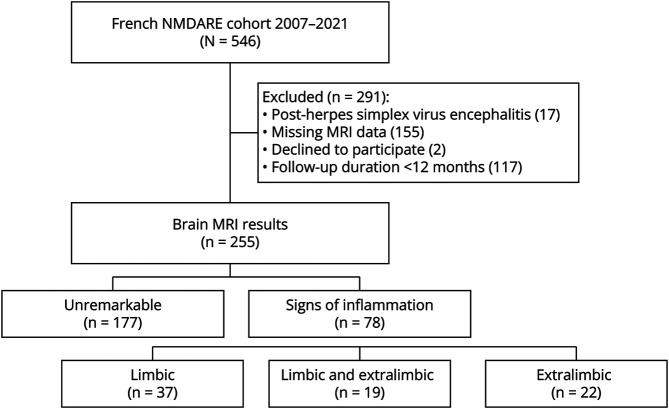
Methods: This retrospective cohort study included patients with NMDARE identified at the French Reference Center for Autoimmune Encephalitis, with at least a one-year follow-up, and with available brain MRI results. In case of brain extralimbic lesion, the image files were reviewed when available. Clinical data were collected from medical records. Multivariable logistic regression analysis was used to study the outcomes at 2-year follow-up; recovery was defined as modified Rankin Scale score ≤1.
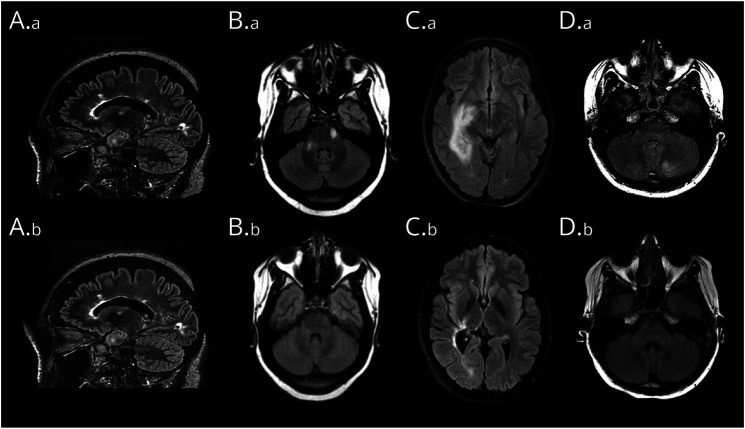
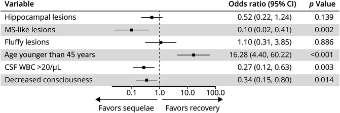
Results: Among the 255 patients included, 37 (14.5%) had limbic hyperintensities and 41 (16.1%) had extralimbic lesions that included multiple sclerosis (MS)-like lesions (14/41, 34.1%); extensive lesions (5/41, 12.2%); and poorly demarcated fluffy lesions, either multifocal (10/41, 24.4%) or involving the cerebral cortex or cerebellum (6/41 each, 14.6%). Extralimbic lesions coexisting with limbic lesions (19/41 patients, 46.3%) were mostly fluffy lesions (11/19, 57.9%). Ten patients had overlapping demyelinating syndromes: 4 with MS, 4 with myelin oligodendrocyte glycoprotein-associated disorder, and 2 with neuromyelitis optica spectrum disorder; all had MS-like (7/10 patients) or extensive (3/10 patients) lesions, and none had fluffy lesions. Extralimbic lesions were associated with symptoms nontypical for NMDARE (23/41, 56.1%, p < 0.001), especially cerebellar ataxia (17/41, 41.5%) and motor impairment (12/41, 29.3%). At 2 years, patients with MS-like or extensive lesions had a lower recovery rate (5/12, 41.7%, and 1/4, 25%, respectively) compared with the patients without extralimbic lesions (124/162, 76.5%; p = 0.014 and p = 0.047, respectively). In multivariable analysis, MS-like lesions, but not hippocampal nor fluffy lesions, were associated with absence of recovery at 2 years (adjusted OR 0.1, 95% CI 0.03-0.42, p = 0.002; extensive lesions [n = 4] not included in the analysis).
Discussion: Brain MRI lesions in NMDARE include limbic hyperintensities and 3 patterns of extralimbic lesions, which are associated with nontypical NMDARE symptoms. Moreover, MS-like and extensive lesions, but not fluffy nor hippocampal lesions, are associated with overlapping demyelinating syndromes and poor clinical outcomes at 2 years. These findings can have practical implications on the monitoring of patients with NMDARE.
期刊介绍:
Neurology Neuroimmunology & Neuroinflammation is an official journal of the American Academy of Neurology. Neurology: Neuroimmunology & Neuroinflammation will be the premier peer-reviewed journal in neuroimmunology and neuroinflammation. This journal publishes rigorously peer-reviewed open-access reports of original research and in-depth reviews of topics in neuroimmunology & neuroinflammation, affecting the full range of neurologic diseases including (but not limited to) Alzheimer's disease, Parkinson's disease, ALS, tauopathy, and stroke; multiple sclerosis and NMO; inflammatory peripheral nerve and muscle disease, Guillain-Barré and myasthenia gravis; nervous system infection; paraneoplastic syndromes, noninfectious encephalitides and other antibody-mediated disorders; and psychiatric and neurodevelopmental disorders. Clinical trials, instructive case reports, and small case series will also be featured.
 求助内容:
求助内容: 应助结果提醒方式:


