Jin Joo Kim, Jin You Kim, Yeon Joo Jeong, Suk Kim, In Sook Lee, Nam Kyung Lee, Taewoo Kang, Heeseung Park, Seokwon Lee
{"title":"Magnetic Resonance Elastography of Invasive Breast Cancer: Evaluating Prognostic Factors and Treatment Response.","authors":"Jin Joo Kim, Jin You Kim, Yeon Joo Jeong, Suk Kim, In Sook Lee, Nam Kyung Lee, Taewoo Kang, Heeseung Park, Seokwon Lee","doi":"10.3390/tomography11020018","DOIUrl":null,"url":null,"abstract":"<p><p><b>Objectives:</b> To assess the elasticity values in breast tissues using magnetic resonance elastography (MRE) and examine the association between elasticity values of invasive breast cancer with prognostic factors and the pathologic response to neoadjuvant systemic therapy (NST). <b>Methods:</b> A total of 57 patients (mean age, 54.1 years) with invasive breast cancers larger than 2 cm in diameter on ultrasound were prospectively enrolled. The elasticity values (mean, minimum, and maximum) of invasive breast cancers, normal fibroglandular tissues, and normal fat tissues were measured via MRE using a commercially available acoustic driver and compared. Elasticity values of breast cancers were compared according to prognostic factors and pathologic responses in patients who received NST before surgery. Receiver operating curve analysis was performed to evaluate the predictive efficacy of elasticity values in terms of pathological response. <b>Results:</b> Among the 57 patients, the mean elasticity value of invasive breast cancers was significantly higher than that of normal fibroglandular tissue and normal fat tissue (7.90 ± 5.80 kPa vs. 2.54 ± 0.80 kPa vs. 1.32 ± 0.33 kPa, all <i>p</i>s < 0.001). Invasive breast cancers with a large diameter (>4 cm) exhibited significantly higher mean elasticity values relative to tumors with a small diameter (≤4 cm) (11.65 ± 7.22 kPa vs. 5.87 ± 3.58 kPa, <i>p</i> = 0.002). Among 24 patients who received NST, mean, minimum, and maximum elasticity values significantly differed between the pathologic complete response (pCR) and non-pCR groups (all <i>p</i>s < 0.05). For the mean elasticity value, the area under the curve value for distinguishing pCR and non-pCR groups was 0.880 (95% confidence interval, 0.682, 0.976; <i>p</i> < 0.001). <b>Conclusions:</b> The elasticity values of invasive breast cancers measured via breast MRE showed a positive correlation with tumor size and showed potential in predicting the therapeutic response in patients receiving NST.</p>","PeriodicalId":51330,"journal":{"name":"Tomography","volume":"11 2","pages":""},"PeriodicalIF":2.2000,"publicationDate":"2025-02-14","publicationTypes":"Journal Article","fieldsOfStudy":null,"isOpenAccess":false,"openAccessPdf":"https://www.ncbi.nlm.nih.gov/pmc/articles/PMC11860845/pdf/","citationCount":"0","resultStr":null,"platform":"Semanticscholar","paperid":null,"PeriodicalName":"Tomography","FirstCategoryId":"3","ListUrlMain":"https://doi.org/10.3390/tomography11020018","RegionNum":4,"RegionCategory":"医学","ArticlePicture":[],"TitleCN":null,"AbstractTextCN":null,"PMCID":null,"EPubDate":"","PubModel":"","JCR":"Q2","JCRName":"RADIOLOGY, NUCLEAR MEDICINE & MEDICAL IMAGING","Score":null,"Total":0}
引用次数: 0
Abstract
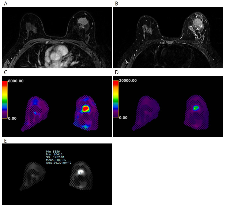
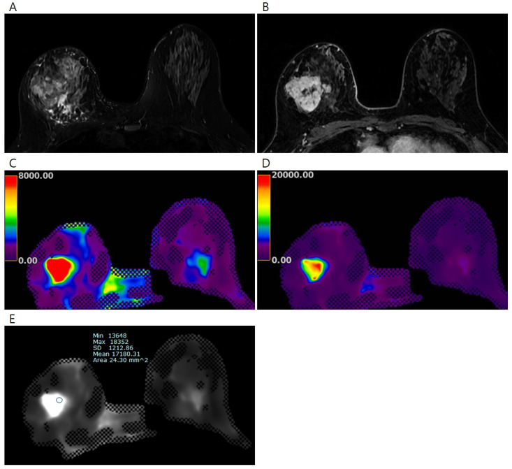
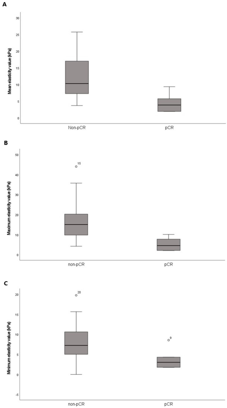
Objectives: To assess the elasticity values in breast tissues using magnetic resonance elastography (MRE) and examine the association between elasticity values of invasive breast cancer with prognostic factors and the pathologic response to neoadjuvant systemic therapy (NST). Methods: A total of 57 patients (mean age, 54.1 years) with invasive breast cancers larger than 2 cm in diameter on ultrasound were prospectively enrolled. The elasticity values (mean, minimum, and maximum) of invasive breast cancers, normal fibroglandular tissues, and normal fat tissues were measured via MRE using a commercially available acoustic driver and compared. Elasticity values of breast cancers were compared according to prognostic factors and pathologic responses in patients who received NST before surgery. Receiver operating curve analysis was performed to evaluate the predictive efficacy of elasticity values in terms of pathological response. Results: Among the 57 patients, the mean elasticity value of invasive breast cancers was significantly higher than that of normal fibroglandular tissue and normal fat tissue (7.90 ± 5.80 kPa vs. 2.54 ± 0.80 kPa vs. 1.32 ± 0.33 kPa, all ps < 0.001). Invasive breast cancers with a large diameter (>4 cm) exhibited significantly higher mean elasticity values relative to tumors with a small diameter (≤4 cm) (11.65 ± 7.22 kPa vs. 5.87 ± 3.58 kPa, p = 0.002). Among 24 patients who received NST, mean, minimum, and maximum elasticity values significantly differed between the pathologic complete response (pCR) and non-pCR groups (all ps < 0.05). For the mean elasticity value, the area under the curve value for distinguishing pCR and non-pCR groups was 0.880 (95% confidence interval, 0.682, 0.976; p < 0.001). Conclusions: The elasticity values of invasive breast cancers measured via breast MRE showed a positive correlation with tumor size and showed potential in predicting the therapeutic response in patients receiving NST.
TomographyMedicine-Radiology, Nuclear Medicine and Imaging
CiteScore
2.70
自引率
10.50%
发文量
222
期刊介绍:
TomographyTM publishes basic (technical and pre-clinical) and clinical scientific articles which involve the advancement of imaging technologies. Tomography encompasses studies that use single or multiple imaging modalities including for example CT, US, PET, SPECT, MR and hyperpolarization technologies, as well as optical modalities (i.e. bioluminescence, photoacoustic, endomicroscopy, fiber optic imaging and optical computed tomography) in basic sciences, engineering, preclinical and clinical medicine.
Tomography also welcomes studies involving exploration and refinement of contrast mechanisms and image-derived metrics within and across modalities toward the development of novel imaging probes for image-based feedback and intervention. The use of imaging in biology and medicine provides unparalleled opportunities to noninvasively interrogate tissues to obtain real-time dynamic and quantitative information required for diagnosis and response to interventions and to follow evolving pathological conditions. As multi-modal studies and the complexities of imaging technologies themselves are ever increasing to provide advanced information to scientists and clinicians.
Tomography provides a unique publication venue allowing investigators the opportunity to more precisely communicate integrated findings related to the diverse and heterogeneous features associated with underlying anatomical, physiological, functional, metabolic and molecular genetic activities of normal and diseased tissue. Thus Tomography publishes peer-reviewed articles which involve the broad use of imaging of any tissue and disease type including both preclinical and clinical investigations. In addition, hardware/software along with chemical and molecular probe advances are welcome as they are deemed to significantly contribute towards the long-term goal of improving the overall impact of imaging on scientific and clinical discovery.

 求助内容:
求助内容: 应助结果提醒方式:


