Julia Gresky, Melina Frotscher, Sophia Thiem, Alexander Stoessel, Alexey Kalmykov, Natalia Berezina
{"title":"Cemento-Osseous Dysplasia in a Female Bronze Age Skeleton (North Caucasus).","authors":"Julia Gresky, Melina Frotscher, Sophia Thiem, Alexander Stoessel, Alexey Kalmykov, Natalia Berezina","doi":"10.1007/s12105-025-01767-1","DOIUrl":null,"url":null,"abstract":"<p><strong>Purpose: </strong>The earliest known case of cemento-osseous dysplasia could be detected in a Bronze Age skeleton, dating back 4500 years ago in the region of the North Caucasus. Although the soft tissue was missing, sufficient diagnosis could be achieved by using different methods that prove the existence of fibro-osseous processes already in prehistory. Skeletal remains provide a direct view of such changes which cannot be obtained from a living patient without compromising.</p><p><strong>Methods: </strong>A skeleton of a 30-40-year-old female individual from the burial mound of Budyonnovsk 10 (including 19 individuals) in Southern Russia was investigated using macroscopic, radiographic, and microscopic methods.</p><p><strong>Results: </strong>In the mandible, destruction of the labial wall of the alveoli 32 and 31 is already visible macroscopically. At the base of the lesion, the original bone is replaced by fine porous bone including small dense particles: plain radiography and computed tomography evidence localized processes to the periapical areas of all lower incisors. The lesions are mainly radiolucent, only the particles in alveolus 32 have a radiopaque appearance. Microscopy shows woven bone as filling of the lesions and additional hypocellular materials in alveolus 32, which can best be explained as cementum-like structures.</p><p><strong>Conclusions: </strong>The lesion´s location in the periapical areas of the lower incisors, the woven bone, and cementum-like structures fit the diagnosis of periapical cemento-osseous dysplasia. The presence of a second individual with focal cemento-osseous dysplasia in this burial mound is an interesting co-occurrence that requires further genetic analysis.</p><p><strong>Limitations: </strong>The diagnosis is solely based on the skeletal remains, soft tissue components are missing.</p><p><strong>Suggestions for further research: </strong>Genetic analyses are planned to detect the underlying mutation for the two individuals.</p>","PeriodicalId":47972,"journal":{"name":"Head & Neck Pathology","volume":"19 1","pages":"28"},"PeriodicalIF":4.1000,"publicationDate":"2025-02-25","publicationTypes":"Journal Article","fieldsOfStudy":null,"isOpenAccess":false,"openAccessPdf":"https://www.ncbi.nlm.nih.gov/pmc/articles/PMC11861816/pdf/","citationCount":"0","resultStr":null,"platform":"Semanticscholar","paperid":null,"PeriodicalName":"Head & Neck Pathology","FirstCategoryId":"1085","ListUrlMain":"https://doi.org/10.1007/s12105-025-01767-1","RegionNum":0,"RegionCategory":null,"ArticlePicture":[],"TitleCN":null,"AbstractTextCN":null,"PMCID":null,"EPubDate":"","PubModel":"","JCR":"Q2","JCRName":"PATHOLOGY","Score":null,"Total":0}
引用次数: 0
Abstract
Purpose: The earliest known case of cemento-osseous dysplasia could be detected in a Bronze Age skeleton, dating back 4500 years ago in the region of the North Caucasus. Although the soft tissue was missing, sufficient diagnosis could be achieved by using different methods that prove the existence of fibro-osseous processes already in prehistory. Skeletal remains provide a direct view of such changes which cannot be obtained from a living patient without compromising.
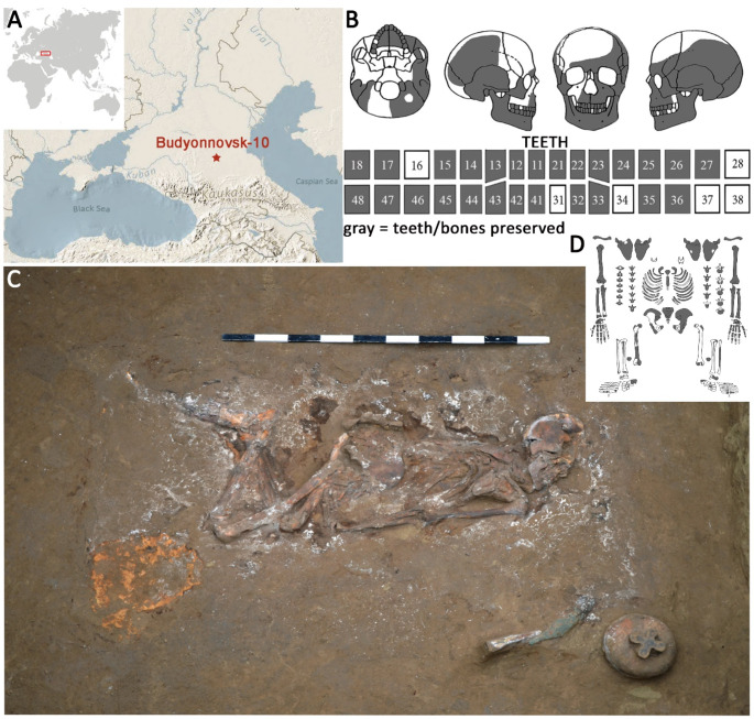
Methods: A skeleton of a 30-40-year-old female individual from the burial mound of Budyonnovsk 10 (including 19 individuals) in Southern Russia was investigated using macroscopic, radiographic, and microscopic methods.
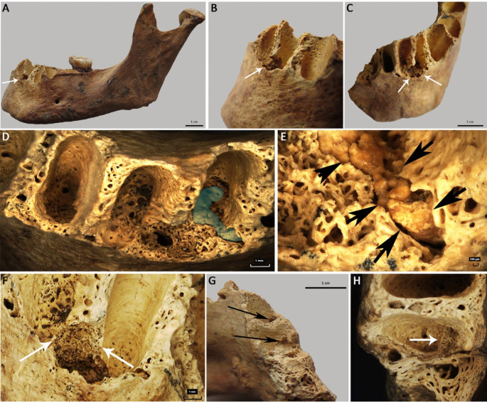
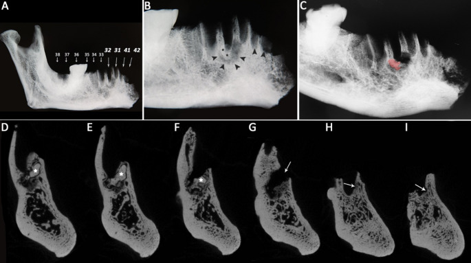
Results: In the mandible, destruction of the labial wall of the alveoli 32 and 31 is already visible macroscopically. At the base of the lesion, the original bone is replaced by fine porous bone including small dense particles: plain radiography and computed tomography evidence localized processes to the periapical areas of all lower incisors. The lesions are mainly radiolucent, only the particles in alveolus 32 have a radiopaque appearance. Microscopy shows woven bone as filling of the lesions and additional hypocellular materials in alveolus 32, which can best be explained as cementum-like structures.
Conclusions: The lesion´s location in the periapical areas of the lower incisors, the woven bone, and cementum-like structures fit the diagnosis of periapical cemento-osseous dysplasia. The presence of a second individual with focal cemento-osseous dysplasia in this burial mound is an interesting co-occurrence that requires further genetic analysis.
Limitations: The diagnosis is solely based on the skeletal remains, soft tissue components are missing.
Suggestions for further research: Genetic analyses are planned to detect the underlying mutation for the two individuals.
期刊介绍:
Head & Neck Pathology presents scholarly papers, reviews and symposia that cover the spectrum of human surgical pathology within the anatomic zones of the oral cavity, sinonasal tract, larynx, hypopharynx, salivary gland, ear and temporal bone, and neck.
The journal publishes rapid developments in new diagnostic criteria, intraoperative consultation, immunohistochemical studies, molecular techniques, genetic analyses, diagnostic aids, experimental pathology, cytology, radiographic imaging, and application of uniform terminology to allow practitioners to continue to maintain and expand their knowledge in the subspecialty of head and neck pathology. Coverage of practical application to daily clinical practice is supported with proceedings and symposia from international societies and academies devoted to this field.
Single-blind peer review
The journal follows a single-blind review procedure, where the reviewers are aware of the names and affiliations of the authors, but the reviewer reports provided to authors are anonymous. Single-blind peer review is the traditional model of peer review that many reviewers are comfortable with, and it facilitates a dispassionate critique of a manuscript.

 求助内容:
求助内容: 应助结果提醒方式:


