Successful resolution of gastric pneumatosis due to a strangulated hiatus hernia and malrotation through non-surgical management: a case report.
IF 0.5
Q4 RADIOLOGY, NUCLEAR MEDICINE & MEDICAL IMAGING
引用次数: 0
Abstract
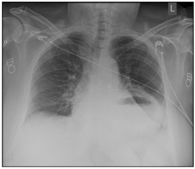
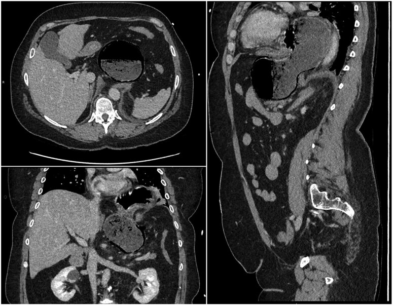
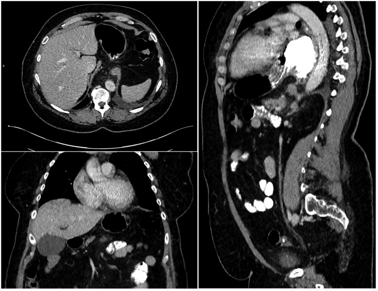
Gastric pneumatosis is a rare finding, and clinicians, when under pressure, find it challenging to immediately identify the cause and decide if the patient requires emergency surgery. We present a case where an initial CT scan demonstrated gastric pneumatosis involving only the greater curvature of the antrum caused by a strangulated hiatus hernia and malrotation of the distal stomach. The CT features suggested the patient required immediate surgery; however, a conservative approach was taken, and a follow-up CT scan 4 days after the onset demonstrated complete resolution and no long-term complications.
通过非手术治疗成功解决绞窄性裂孔疝和旋转不良引起的胃肺病1例报告。
胃肺病是一种罕见的发现,临床医生在压力下发现立即确定原因并决定患者是否需要紧急手术是具有挑战性的。我们提出了一个病例,最初的CT扫描显示胃肺病仅涉及由绞窄性裂孔疝和远端胃旋转不良引起的胃窦较大弯曲。CT表现提示患者需要立即手术;然而,我们采取了保守的方法,发病4天后的随访CT扫描显示完全消退,没有长期并发症。
本文章由计算机程序翻译,如有差异,请以英文原文为准。
求助全文
约1分钟内获得全文
求助全文

 求助内容:
求助内容: 应助结果提醒方式:


