{"title":"Investigation of Antibody Pharmacokinetics in Male Reproductive System and Its Characterization Using a Translational PBPK Model.","authors":"Sree Ojili, Dhaval K Shah","doi":"10.3390/antib14010017","DOIUrl":null,"url":null,"abstract":"<p><p><b>Objectives:</b> To investigate the pharmacokinetics (PK) of the monoclonal antibody (mAb) in male reproductive tissues and develop a translational physiologically based pharmacokinetic (PBPK) model to characterize the PK data. <b>Method:</b> The PK of a non-cross-reactive antibody (trastuzumab) was investigated in human FcRn-expressing male mice following a 10 mg/kg intravenous dose. The PK in plasma and male reproductive tissues (i.e., epididymis, testes, vas deferens, seminal vesicles, and prostate glands) were evaluated. The observed PK data in mice were mathematically characterized using a novel PBPK model for antibodies that contained male reproductive systems. The mouse PBPK model was scaled to rats, monkeys, and humans to predict the PK of antibodies in male reproductive organs across animal species. <b>Results</b>: Plasma and tissue PK data generated in mice suggest that antibody distribution in male reproductive tissues is generally lower compared to that of most of the organs. The antibody exposure in the testes was 1.70%, in the epididymis was 2.57%, in the vas deferens was 2.01%, in the seminal vesicle was 0.42%, and in the prostate gland was 0.52% of the plasma exposure. The plasma and tissue PK data were simultaneously characterized using the PBPK model, which incorporated the novel male reproductive system. All the predicted PK profiles were within two-fold of the observed data, as indicated by percentage prediction error (%PE) values. The mouse model was successfully translated to bigger animals, and the model was used to simulate the PK of antibodies in rat, monkey, and human male reproductive systems. <b>Conclusions</b>: The combination of the experimental data and novel PBPK model presented here provides unprecedented insights into the antibody distributions in different male reproductive tissues. The PBPK model can serve as a crucial tool for advancing the development of antibody-based therapies for treating sexually transmitted infections (STIs), cancers, and contraceptives.</p>","PeriodicalId":8188,"journal":{"name":"Antibodies","volume":"14 1","pages":""},"PeriodicalIF":2.7000,"publicationDate":"2025-02-13","publicationTypes":"Journal Article","fieldsOfStudy":null,"isOpenAccess":false,"openAccessPdf":"https://www.ncbi.nlm.nih.gov/pmc/articles/PMC11843977/pdf/","citationCount":"0","resultStr":null,"platform":"Semanticscholar","paperid":null,"PeriodicalName":"Antibodies","FirstCategoryId":"1085","ListUrlMain":"https://doi.org/10.3390/antib14010017","RegionNum":0,"RegionCategory":null,"ArticlePicture":[],"TitleCN":null,"AbstractTextCN":null,"PMCID":null,"EPubDate":"","PubModel":"","JCR":"Q3","JCRName":"IMMUNOLOGY","Score":null,"Total":0}
引用次数: 0
Abstract
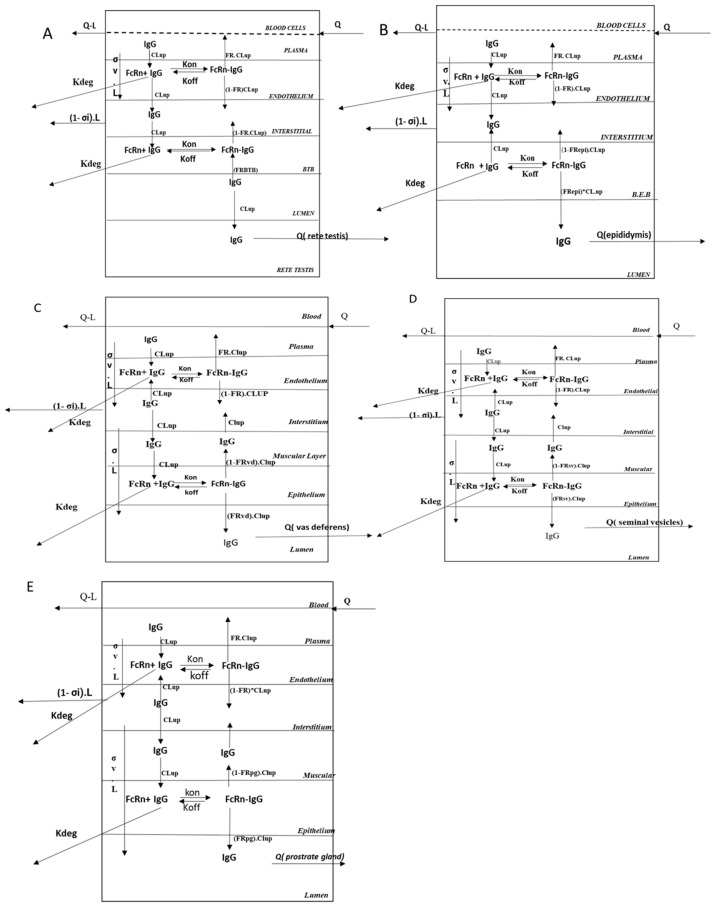
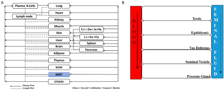
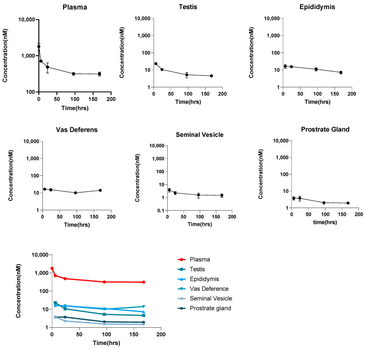
Objectives: To investigate the pharmacokinetics (PK) of the monoclonal antibody (mAb) in male reproductive tissues and develop a translational physiologically based pharmacokinetic (PBPK) model to characterize the PK data. Method: The PK of a non-cross-reactive antibody (trastuzumab) was investigated in human FcRn-expressing male mice following a 10 mg/kg intravenous dose. The PK in plasma and male reproductive tissues (i.e., epididymis, testes, vas deferens, seminal vesicles, and prostate glands) were evaluated. The observed PK data in mice were mathematically characterized using a novel PBPK model for antibodies that contained male reproductive systems. The mouse PBPK model was scaled to rats, monkeys, and humans to predict the PK of antibodies in male reproductive organs across animal species. Results: Plasma and tissue PK data generated in mice suggest that antibody distribution in male reproductive tissues is generally lower compared to that of most of the organs. The antibody exposure in the testes was 1.70%, in the epididymis was 2.57%, in the vas deferens was 2.01%, in the seminal vesicle was 0.42%, and in the prostate gland was 0.52% of the plasma exposure. The plasma and tissue PK data were simultaneously characterized using the PBPK model, which incorporated the novel male reproductive system. All the predicted PK profiles were within two-fold of the observed data, as indicated by percentage prediction error (%PE) values. The mouse model was successfully translated to bigger animals, and the model was used to simulate the PK of antibodies in rat, monkey, and human male reproductive systems. Conclusions: The combination of the experimental data and novel PBPK model presented here provides unprecedented insights into the antibody distributions in different male reproductive tissues. The PBPK model can serve as a crucial tool for advancing the development of antibody-based therapies for treating sexually transmitted infections (STIs), cancers, and contraceptives.
期刊介绍:
Antibodies (ISSN 2073-4468), an international, peer-reviewed open access journal which provides an advanced forum for studies related to antibodies and antigens. It publishes reviews, research articles, communications and short notes. Our aim is to encourage scientists to publish their experimental and theoretical results in as much detail as possible. There is no restriction on the length of the papers. Full experimental and/or methodical details must be provided. Electronic files or software regarding the full details of the calculation and experimental procedure - if unable to be published in a normal way - can be deposited as supplementary material. This journal covers all topics related to antibodies and antigens, topics of interest include (but are not limited to): antibody-producing cells (including B cells), antibody structure and function, antibody-antigen interactions, Fc receptors, antibody manufacturing antibody engineering, antibody therapy, immunoassays, antibody diagnosis, tissue antigens, exogenous antigens, endogenous antigens, autoantigens, monoclonal antibodies, natural antibodies, humoral immune responses, immunoregulatory molecules.

 求助内容:
求助内容: 应助结果提醒方式:


