P Sanjay, Vittal Manohar, Sushmita Balol, Yashwanth M B Naik
{"title":"Diagnostic performance of contrast-enhanced T2-FLAIR MRI in the detection of meningitis.","authors":"P Sanjay, Vittal Manohar, Sushmita Balol, Yashwanth M B Naik","doi":"10.4102/sajr.v29i1.3018","DOIUrl":null,"url":null,"abstract":"<p><strong>Background: </strong>The contrast-enhanced T2-FLAIR (CE-T2-FLAIR) sequence on MRI, through the suppression of CSF and vascular signals, can detect subtle meningeal enhancement in meningitis that may not be appreciable on the routinely used contrast-enhanced T1W (CE-T1W) sequence.</p><p><strong>Objectives: </strong>To assess CE-T2-FLAIR compared to CE-T1W in the diagnosis of meningitis, using CSF analysis as the gold standard, using both qualitative and quantitative approaches for assessment.</p><p><strong>Method: </strong>A retrospective study was conducted on 53 patients with clinically suspected meningitis referred for brain MRI. Twenty-seven patients, positive for meningitis on CSF analysis, were classified as the case group; the remaining patients were designated as controls. The pre-contrast, CE-T1W and CE-T2-FLAIR images were assessed and analysed, qualitatively for the detection of abnormal meningeal enhancement, and quantitatively by measuring single pixel signal intensities (SPSI) over the meninges and vessels.</p><p><strong>Results: </strong>Contrast-enhanced T2-FLAIR demonstrated significantly higher sensitivity (92.59% vs. 57.69%), negative predictive value (92.59% vs. 70.27%) and diagnostic accuracy (94.34% vs. 78.85%) compared to CE-T1W. Additionally, CE-T2-FLAIR showed significantly greater meningeal SPSI and enhancement than CE-T1W.</p><p><strong>Conclusion: </strong>Contrast-enhanced T2-FLAIR is better at detecting abnormal meningeal enhancement in meningitis than CE-T1W, because of significantly greater signal intensity and enhancement of the meninges compared to vessels.</p><p><strong>Contribution: </strong>This study reiterates the usefulness of CE-T2-FLAIR as an additional sequence for the detection of abnormal meningeal enhancement in cases of meningitis as confirmed both qualitatively and quantitatively.</p>","PeriodicalId":43442,"journal":{"name":"SA Journal of Radiology","volume":"29 1","pages":"3018"},"PeriodicalIF":0.9000,"publicationDate":"2025-01-27","publicationTypes":"Journal Article","fieldsOfStudy":null,"isOpenAccess":false,"openAccessPdf":"https://www.ncbi.nlm.nih.gov/pmc/articles/PMC11830885/pdf/","citationCount":"0","resultStr":null,"platform":"Semanticscholar","paperid":null,"PeriodicalName":"SA Journal of Radiology","FirstCategoryId":"1085","ListUrlMain":"https://doi.org/10.4102/sajr.v29i1.3018","RegionNum":0,"RegionCategory":null,"ArticlePicture":[],"TitleCN":null,"AbstractTextCN":null,"PMCID":null,"EPubDate":"2025/1/1 0:00:00","PubModel":"eCollection","JCR":"Q4","JCRName":"RADIOLOGY, NUCLEAR MEDICINE & MEDICAL IMAGING","Score":null,"Total":0}
引用次数: 0
Abstract
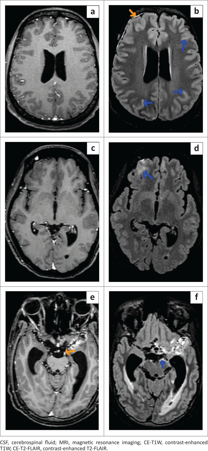
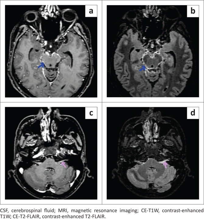
Background: The contrast-enhanced T2-FLAIR (CE-T2-FLAIR) sequence on MRI, through the suppression of CSF and vascular signals, can detect subtle meningeal enhancement in meningitis that may not be appreciable on the routinely used contrast-enhanced T1W (CE-T1W) sequence.
Objectives: To assess CE-T2-FLAIR compared to CE-T1W in the diagnosis of meningitis, using CSF analysis as the gold standard, using both qualitative and quantitative approaches for assessment.
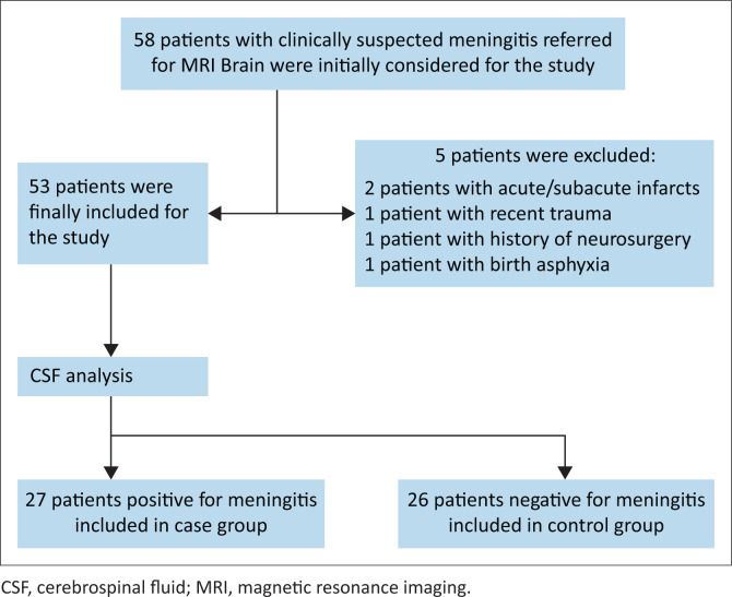
Method: A retrospective study was conducted on 53 patients with clinically suspected meningitis referred for brain MRI. Twenty-seven patients, positive for meningitis on CSF analysis, were classified as the case group; the remaining patients were designated as controls. The pre-contrast, CE-T1W and CE-T2-FLAIR images were assessed and analysed, qualitatively for the detection of abnormal meningeal enhancement, and quantitatively by measuring single pixel signal intensities (SPSI) over the meninges and vessels.
Results: Contrast-enhanced T2-FLAIR demonstrated significantly higher sensitivity (92.59% vs. 57.69%), negative predictive value (92.59% vs. 70.27%) and diagnostic accuracy (94.34% vs. 78.85%) compared to CE-T1W. Additionally, CE-T2-FLAIR showed significantly greater meningeal SPSI and enhancement than CE-T1W.
Conclusion: Contrast-enhanced T2-FLAIR is better at detecting abnormal meningeal enhancement in meningitis than CE-T1W, because of significantly greater signal intensity and enhancement of the meninges compared to vessels.
Contribution: This study reiterates the usefulness of CE-T2-FLAIR as an additional sequence for the detection of abnormal meningeal enhancement in cases of meningitis as confirmed both qualitatively and quantitatively.
背景:MRI上的对比增强T2-FLAIR (CE-T2-FLAIR)序列,通过抑制脑脊液和血管信号,可以检测到脑膜炎中细微的脑膜强化,而常规使用的对比增强T1W (CE-T1W)序列可能无法察觉。目的:评价CE-T2-FLAIR与CE-T1W对脑膜炎的诊断价值,以脑脊液分析为金标准,采用定性和定量方法进行评估。方法:对53例临床疑似脑膜炎患者行脑MRI检查进行回顾性分析。27例脑脊液分析呈脑膜炎阳性的患者为病例组;其余患者被指定为对照组。对对比前、CE-T1W和CE-T2-FLAIR图像进行评估和分析,定性地检测异常脑膜增强,定量地测量脑膜和血管上的单像素信号强度(SPSI)。结果:对比增强T2-FLAIR的敏感性(92.59% vs. 57.69%)、阴性预测值(92.59% vs. 70.27%)和诊断准确率(94.34% vs. 78.85%)均明显高于CE-T1W。此外,CE-T2-FLAIR比CE-T1W显示更大的脑膜SPSI和增强。结论:对比增强T2-FLAIR比CE-T1W更能发现脑膜炎的异常脑膜强化,其信号强度和强化程度均明显高于血管。贡献:本研究重申了CE-T2-FLAIR作为检测脑膜炎病例异常脑膜增强的额外序列的有效性,这在定性和定量上都得到了证实。
期刊介绍:
The SA Journal of Radiology is the official journal of the Radiological Society of South Africa and the Professional Association of Radiologists in South Africa and Namibia. The SA Journal of Radiology is a general diagnostic radiological journal which carries original research and review articles, pictorial essays, case reports, letters, editorials, radiological practice and other radiological articles.

 求助内容:
求助内容: 应助结果提醒方式:


