Caliper verification and gap measurements of kinematic alignment total knee arthroplasty utilizing an imageless, accelerometer-based navigation system.
James H Sikes, Drew P Melancon, Isaac J Spears, Evan H Powers, Spencer J Montgomery
{"title":"Caliper verification and gap measurements of kinematic alignment total knee arthroplasty utilizing an imageless, accelerometer-based navigation system.","authors":"James H Sikes, Drew P Melancon, Isaac J Spears, Evan H Powers, Spencer J Montgomery","doi":"10.1186/s43019-025-00260-x","DOIUrl":null,"url":null,"abstract":"<p><strong>Purpose: </strong>Kinematic alignment (KA) in total knee arthroplasty (TKA) aims to restore the patient's knee to the prearthritic state. The purpose of this study was to investigate the accuracy of using an implant-agnostic, imageless, accelerometer-based navigation system to perform KA TKA on the basis of caliper verification and quantification of the flexion and extension gaps.</p><p><strong>Materials and methods: </strong>Seven cadaveric lower extremities underwent primary TKA utilizing a kinematic alignment workflow with the imageless navigation system. Accuracy of the technique was confirmed through caliper verification of bone cuts.</p><p><strong>Results: </strong>All cuts were within 1 mm of anticipated measurements, except for the lateral tibial fragment, which averaged 1 mm (standard deviation [SD] 0.9 mm) thicker than anticipated. In extension, medial and lateral gaps were symmetric and averaged within 0.6 mm of expectation. In flexion, the medial gap averaged within 0.5 mm of expectation, while the lateral gap averaged 2.6 mm larger than the symmetric expectation, consistently producing a trapezoidal space.</p><p><strong>Conclusions: </strong>The implementation of an accelerometer-based navigation system in KA TKA allows for highly accurate results, which was confirmed with caliper verification. This workflow produced a symmetric extension gap and a trapezoidal flexion gap with an average increased lateral flexion gap of 2.6 mm compared with the medial side.</p>","PeriodicalId":36317,"journal":{"name":"Knee Surgery and Related Research","volume":"37 1","pages":"8"},"PeriodicalIF":4.4000,"publicationDate":"2025-02-17","publicationTypes":"Journal Article","fieldsOfStudy":null,"isOpenAccess":false,"openAccessPdf":"https://www.ncbi.nlm.nih.gov/pmc/articles/PMC11834236/pdf/","citationCount":"0","resultStr":null,"platform":"Semanticscholar","paperid":null,"PeriodicalName":"Knee Surgery and Related Research","FirstCategoryId":"1085","ListUrlMain":"https://doi.org/10.1186/s43019-025-00260-x","RegionNum":0,"RegionCategory":null,"ArticlePicture":[],"TitleCN":null,"AbstractTextCN":null,"PMCID":null,"EPubDate":"","PubModel":"","JCR":"Q2","JCRName":"Medicine","Score":null,"Total":0}
引用次数: 0
Abstract
Purpose: Kinematic alignment (KA) in total knee arthroplasty (TKA) aims to restore the patient's knee to the prearthritic state. The purpose of this study was to investigate the accuracy of using an implant-agnostic, imageless, accelerometer-based navigation system to perform KA TKA on the basis of caliper verification and quantification of the flexion and extension gaps.
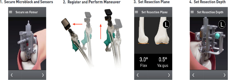
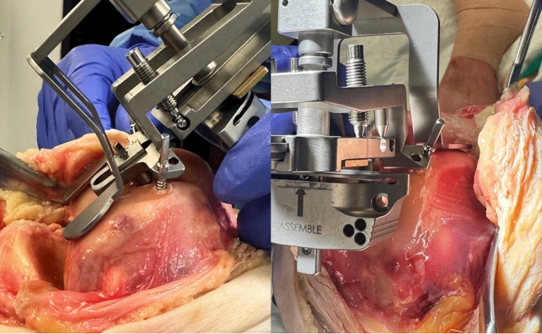
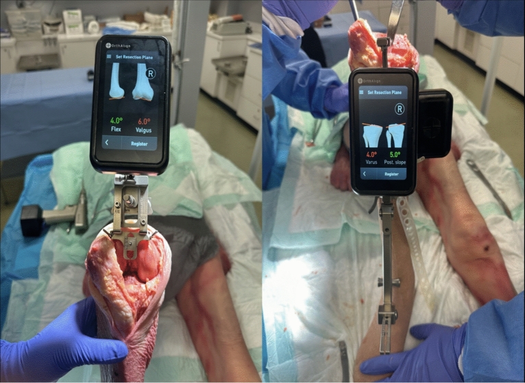
Materials and methods: Seven cadaveric lower extremities underwent primary TKA utilizing a kinematic alignment workflow with the imageless navigation system. Accuracy of the technique was confirmed through caliper verification of bone cuts.
Results: All cuts were within 1 mm of anticipated measurements, except for the lateral tibial fragment, which averaged 1 mm (standard deviation [SD] 0.9 mm) thicker than anticipated. In extension, medial and lateral gaps were symmetric and averaged within 0.6 mm of expectation. In flexion, the medial gap averaged within 0.5 mm of expectation, while the lateral gap averaged 2.6 mm larger than the symmetric expectation, consistently producing a trapezoidal space.
Conclusions: The implementation of an accelerometer-based navigation system in KA TKA allows for highly accurate results, which was confirmed with caliper verification. This workflow produced a symmetric extension gap and a trapezoidal flexion gap with an average increased lateral flexion gap of 2.6 mm compared with the medial side.

 求助内容:
求助内容: 应助结果提醒方式:


