Cristian Niky Cumpătă, Maria Cristina Munteanu, Elena Cristina Andrei, Ilona Mihaela Liliac, Cristina Jana Busuioc, Paolo Di Francesco, Mădălina Anca Moldovan, Simona Iuliana Enache, Alexandru Burcea, Ciprian Laurenţiu Pătru, Călin Rareş Roman
{"title":"Comprehensive insights into Pindborg tumor: etiology, advanced diagnostic approaches, and evidence-based management strategies - review of literature.","authors":"Cristian Niky Cumpătă, Maria Cristina Munteanu, Elena Cristina Andrei, Ilona Mihaela Liliac, Cristina Jana Busuioc, Paolo Di Francesco, Mădălina Anca Moldovan, Simona Iuliana Enache, Alexandru Burcea, Ciprian Laurenţiu Pătru, Călin Rareş Roman","doi":"10.47162/RJME.65.4.08","DOIUrl":null,"url":null,"abstract":"<p><p>Pindborg tumor is a calcifying epithelial odontogenic tumor possibly arising from developmental disturbances in dental lamina remnants. It predominantly affects individuals in their third decade of life, with women also experiencing later onset. The tumor exists in two forms, namely intraosseous (central) and extraosseous (peripheral), with the former showing higher post-surgery recurrence rates of about 14%. Despite its rarity, the tumor can be misdiagnosed due to symptoms resembling dental issues and headaches, or it may even be asymptomatic. Radiologically, it presents a mix of radiolucent and radiopaque areas, sometimes unilocular or multilocular. Histopathologically, it is characterized by nests and sheets of polygonal epithelial cells with eosinophilic cytoplasm and prominent nucleoli. The presence of eosinophilic amyloid-like material and calcifications is distinctive, ranging from small concretions to larger aggregates. The exact origin of amyloids is unknown, but they are thought to derive from degraded keratin filaments. Treatment varies by tumor location, with more invasive procedures required for jaw tumors, including bone resection, due to their aggressive growth and invasion of the surrounding tissues. Accurate, individualized treatment is crucial for patient outcomes, particularly in cases where the tumor's calcification is absent, indicating a severe impact on health. Our study included a case report of a 12-year-old patient who presented to the dental clinic complaining of sporadic pain in the area of the lower right front teeth. During a clinical examination of the area, we noticed a deformation of the alveolar bone, with a depressed mucosa. We followed the chronological steps of radiological examination, lesion excision, and histopathological examination to obtain a definitive diagnosis.</p>","PeriodicalId":54447,"journal":{"name":"Romanian Journal of Morphology and Embryology","volume":"65 4","pages":"617-625"},"PeriodicalIF":1.5000,"publicationDate":"2024-10-01","publicationTypes":"Journal Article","fieldsOfStudy":null,"isOpenAccess":false,"openAccessPdf":"https://www.ncbi.nlm.nih.gov/pmc/articles/PMC11924921/pdf/","citationCount":"0","resultStr":null,"platform":"Semanticscholar","paperid":null,"PeriodicalName":"Romanian Journal of Morphology and Embryology","FirstCategoryId":"3","ListUrlMain":"https://doi.org/10.47162/RJME.65.4.08","RegionNum":4,"RegionCategory":"医学","ArticlePicture":[],"TitleCN":null,"AbstractTextCN":null,"PMCID":null,"EPubDate":"","PubModel":"","JCR":"Q4","JCRName":"DEVELOPMENTAL BIOLOGY","Score":null,"Total":0}
引用次数: 0
Abstract
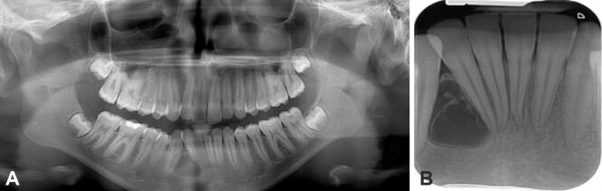
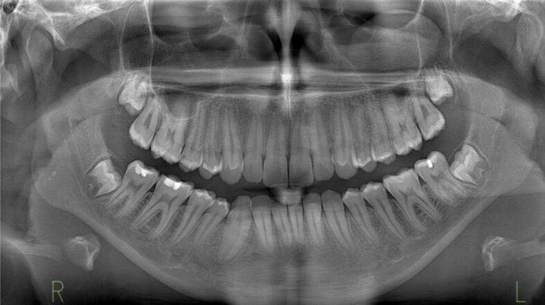
Pindborg tumor is a calcifying epithelial odontogenic tumor possibly arising from developmental disturbances in dental lamina remnants. It predominantly affects individuals in their third decade of life, with women also experiencing later onset. The tumor exists in two forms, namely intraosseous (central) and extraosseous (peripheral), with the former showing higher post-surgery recurrence rates of about 14%. Despite its rarity, the tumor can be misdiagnosed due to symptoms resembling dental issues and headaches, or it may even be asymptomatic. Radiologically, it presents a mix of radiolucent and radiopaque areas, sometimes unilocular or multilocular. Histopathologically, it is characterized by nests and sheets of polygonal epithelial cells with eosinophilic cytoplasm and prominent nucleoli. The presence of eosinophilic amyloid-like material and calcifications is distinctive, ranging from small concretions to larger aggregates. The exact origin of amyloids is unknown, but they are thought to derive from degraded keratin filaments. Treatment varies by tumor location, with more invasive procedures required for jaw tumors, including bone resection, due to their aggressive growth and invasion of the surrounding tissues. Accurate, individualized treatment is crucial for patient outcomes, particularly in cases where the tumor's calcification is absent, indicating a severe impact on health. Our study included a case report of a 12-year-old patient who presented to the dental clinic complaining of sporadic pain in the area of the lower right front teeth. During a clinical examination of the area, we noticed a deformation of the alveolar bone, with a depressed mucosa. We followed the chronological steps of radiological examination, lesion excision, and histopathological examination to obtain a definitive diagnosis.
期刊介绍:
Romanian Journal of Morphology and Embryology (Rom J Morphol Embryol) publishes studies on all aspects of normal morphology and human comparative and experimental pathology. The Journal accepts only researches that utilize modern investigation methods (studies of anatomy, pathology, cytopathology, immunohistochemistry, histochemistry, immunology, morphometry, molecular and cellular biology, electronic microscopy, etc.).

 求助内容:
求助内容: 应助结果提醒方式:


