Nicholas Senn, P James Ross, Reina Ayde, Vasiliki Mallikourti, Adarsh Krishna, Charly James, Clarisse F de Vries, Lionel M Broche, Gordon D Waiter, Mary Joan MacLeod
{"title":"Field-cycling imaging yields repeatable brain R<sub>1</sub> dispersion measurement at fields strengths below 0.2 Tesla with optimal fitting routine.","authors":"Nicholas Senn, P James Ross, Reina Ayde, Vasiliki Mallikourti, Adarsh Krishna, Charly James, Clarisse F de Vries, Lionel M Broche, Gordon D Waiter, Mary Joan MacLeod","doi":"10.1007/s10334-025-01230-w","DOIUrl":null,"url":null,"abstract":"<p><strong>Objectives: </strong>By rapidly changing magnetic field strength between 0.2 and 200 mT during the pulse sequence Field-Cycling Imaging (FCI) makes it possible to identify and evaluate new quantitative markers of pathology derived from dispersion of spin-lattice relaxation rate (R<sub>1</sub>) in vivo. The aim of this work was to determine the most effective approach to reliably estimate multi-field R<sub>1</sub> dispersion measurements in brain tissue using FCI.</p><p><strong>Materials and methods: </strong>This repeatability study consisted of twenty participants with moderate or severe small vessel disease. Each participant underwent 3 T MRI and FCI scans, repeated 30 days apart. After R<sub>1</sub> maps were generated at 0.2, 2, 20, and 200 mT, co-registered tissue labels generated from 3 T MRI were used to extract tissue averaged values of R<sub>1</sub> dispersion from regions of white matter (WM) and WM hyperintensities (WMHs).</p><p><strong>Results: </strong>The fitted model which yielded best overall image contrast between WM and WMH regions and R<sub>1</sub> dispersion model adherence was determined. Tissue averaged values of R<sub>1</sub> (0.2 mT) and R<sub>1</sub> dispersion slope exhibited Cohen's d effect sizes of 3.07 and 1.48, respectively, between regions of WM and WMH. The cohort study results were repeatable between study visits.</p><p><strong>Discussion: </strong>Differences in R<sub>1</sub> measurements could repeatably be discerned between normal and abnormal appearing brain tissues.</p>","PeriodicalId":18067,"journal":{"name":"Magnetic Resonance Materials in Physics, Biology and Medicine","volume":" ","pages":"465-474"},"PeriodicalIF":2.5000,"publicationDate":"2025-07-01","publicationTypes":"Journal Article","fieldsOfStudy":null,"isOpenAccess":false,"openAccessPdf":"https://www.ncbi.nlm.nih.gov/pmc/articles/PMC12255585/pdf/","citationCount":"0","resultStr":null,"platform":"Semanticscholar","paperid":null,"PeriodicalName":"Magnetic Resonance Materials in Physics, Biology and Medicine","FirstCategoryId":"3","ListUrlMain":"https://doi.org/10.1007/s10334-025-01230-w","RegionNum":4,"RegionCategory":"医学","ArticlePicture":[],"TitleCN":null,"AbstractTextCN":null,"PMCID":null,"EPubDate":"2025/2/15 0:00:00","PubModel":"Epub","JCR":"Q3","JCRName":"RADIOLOGY, NUCLEAR MEDICINE & MEDICAL IMAGING","Score":null,"Total":0}
引用次数: 0
Abstract
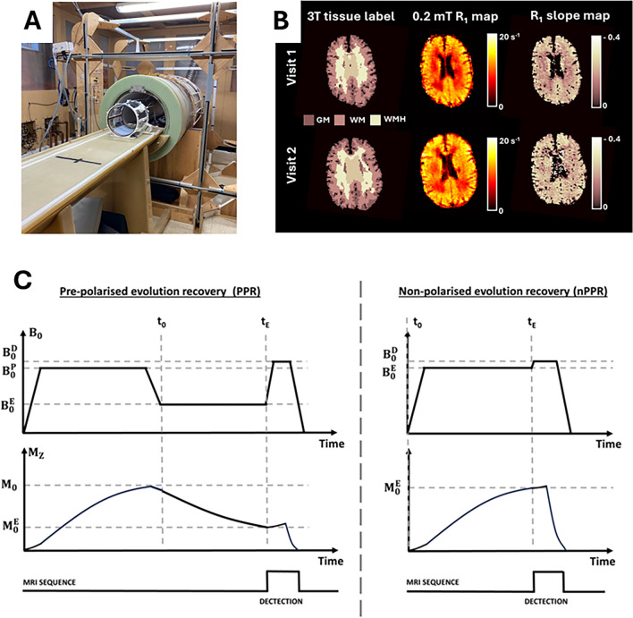
Objectives: By rapidly changing magnetic field strength between 0.2 and 200 mT during the pulse sequence Field-Cycling Imaging (FCI) makes it possible to identify and evaluate new quantitative markers of pathology derived from dispersion of spin-lattice relaxation rate (R1) in vivo. The aim of this work was to determine the most effective approach to reliably estimate multi-field R1 dispersion measurements in brain tissue using FCI.
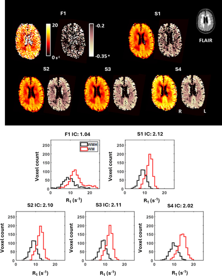
Materials and methods: This repeatability study consisted of twenty participants with moderate or severe small vessel disease. Each participant underwent 3 T MRI and FCI scans, repeated 30 days apart. After R1 maps were generated at 0.2, 2, 20, and 200 mT, co-registered tissue labels generated from 3 T MRI were used to extract tissue averaged values of R1 dispersion from regions of white matter (WM) and WM hyperintensities (WMHs).
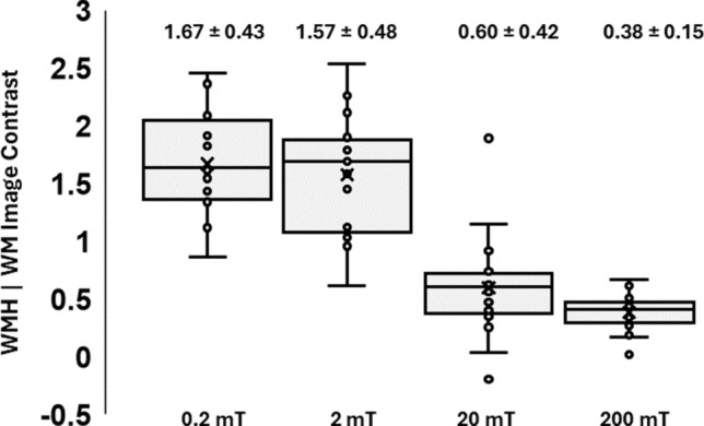
Results: The fitted model which yielded best overall image contrast between WM and WMH regions and R1 dispersion model adherence was determined. Tissue averaged values of R1 (0.2 mT) and R1 dispersion slope exhibited Cohen's d effect sizes of 3.07 and 1.48, respectively, between regions of WM and WMH. The cohort study results were repeatable between study visits.
Discussion: Differences in R1 measurements could repeatably be discerned between normal and abnormal appearing brain tissues.
期刊介绍:
MAGMA is a multidisciplinary international journal devoted to the publication of articles on all aspects of magnetic resonance techniques and their applications in medicine and biology. MAGMA currently publishes research papers, reviews, letters to the editor, and commentaries, six times a year. The subject areas covered by MAGMA include:
advances in materials, hardware and software in magnetic resonance technology,
new developments and results in research and practical applications of magnetic resonance imaging and spectroscopy related to biology and medicine,
study of animal models and intact cells using magnetic resonance,
reports of clinical trials on humans and clinical validation of magnetic resonance protocols.

 求助内容:
求助内容: 应助结果提醒方式:


