{"title":"A method to measure renal inner medullary perfusion using MR renography.","authors":"A de Boer, K Sharma, B Alhummiany, S P Sourbron","doi":"10.1007/s10334-025-01225-7","DOIUrl":null,"url":null,"abstract":"<p><strong>Objective: </strong>In the kidney, the medulla is most susceptible to damage in case of hampered perfusion or oxygenation. Due to separate regulation of cortical and medullary perfusion, measurement of both is crucial to improve the understanding of renal pathophysiology. We aim to develop and evaluate a physiologically accurate model to measure renal inner medullary (F<sub>med</sub>) and cortical perfusion (F<sub>cor</sub>) separately.</p><p><strong>Materials and methods: </strong>We developed a 7-compartment model of renal perfusion and used an iterated approach to fit 10 free parameters. Model stability and accuracy were tested on both patient data and simulations. Cortical perfusion and F<sub>T</sub> (tubular flow or glomerular filtration rate per unit of tissue volume) were compared to a conventional 2-compartment filtration model.</p><p><strong>Results: </strong>Average (standard deviation) F<sub>med</sub> was 37(23)mL/100 mL/min. Fitting stability as expressed by the median (interquartile range) coefficient of variation between fits was 0.0(0.0-5.8)%, with outliers up to 81%. In simulations, F<sub>med</sub> was underestimated by around 8%. Intra-class correlation coefficients for F<sub>cor</sub> and F<sub>T</sub> as measured with the 2- and 7- compartment model were 0.87 and 0.63, respectively.</p><p><strong>Discussion: </strong>We developed a pharmacokinetic model closely following renal physiology. Although the results were vulnerable for overfitting, relatively stable results could be obtained even for F<sub>med</sub>.</p>","PeriodicalId":18067,"journal":{"name":"Magnetic Resonance Materials in Physics, Biology and Medicine","volume":" ","pages":"791-802"},"PeriodicalIF":2.5000,"publicationDate":"2025-10-01","publicationTypes":"Journal Article","fieldsOfStudy":null,"isOpenAccess":false,"openAccessPdf":"https://www.ncbi.nlm.nih.gov/pmc/articles/PMC12497669/pdf/","citationCount":"0","resultStr":null,"platform":"Semanticscholar","paperid":null,"PeriodicalName":"Magnetic Resonance Materials in Physics, Biology and Medicine","FirstCategoryId":"3","ListUrlMain":"https://doi.org/10.1007/s10334-025-01225-7","RegionNum":4,"RegionCategory":"医学","ArticlePicture":[],"TitleCN":null,"AbstractTextCN":null,"PMCID":null,"EPubDate":"2025/2/15 0:00:00","PubModel":"Epub","JCR":"Q3","JCRName":"RADIOLOGY, NUCLEAR MEDICINE & MEDICAL IMAGING","Score":null,"Total":0}
引用次数: 0
Abstract
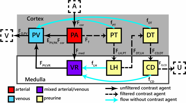
Objective: In the kidney, the medulla is most susceptible to damage in case of hampered perfusion or oxygenation. Due to separate regulation of cortical and medullary perfusion, measurement of both is crucial to improve the understanding of renal pathophysiology. We aim to develop and evaluate a physiologically accurate model to measure renal inner medullary (Fmed) and cortical perfusion (Fcor) separately.
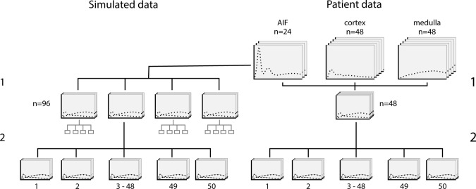
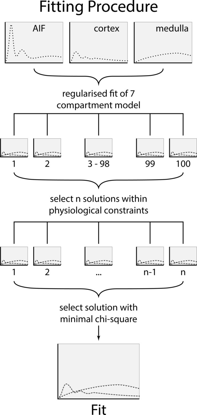
Materials and methods: We developed a 7-compartment model of renal perfusion and used an iterated approach to fit 10 free parameters. Model stability and accuracy were tested on both patient data and simulations. Cortical perfusion and FT (tubular flow or glomerular filtration rate per unit of tissue volume) were compared to a conventional 2-compartment filtration model.
Results: Average (standard deviation) Fmed was 37(23)mL/100 mL/min. Fitting stability as expressed by the median (interquartile range) coefficient of variation between fits was 0.0(0.0-5.8)%, with outliers up to 81%. In simulations, Fmed was underestimated by around 8%. Intra-class correlation coefficients for Fcor and FT as measured with the 2- and 7- compartment model were 0.87 and 0.63, respectively.
Discussion: We developed a pharmacokinetic model closely following renal physiology. Although the results were vulnerable for overfitting, relatively stable results could be obtained even for Fmed.
期刊介绍:
MAGMA is a multidisciplinary international journal devoted to the publication of articles on all aspects of magnetic resonance techniques and their applications in medicine and biology. MAGMA currently publishes research papers, reviews, letters to the editor, and commentaries, six times a year. The subject areas covered by MAGMA include:
advances in materials, hardware and software in magnetic resonance technology,
new developments and results in research and practical applications of magnetic resonance imaging and spectroscopy related to biology and medicine,
study of animal models and intact cells using magnetic resonance,
reports of clinical trials on humans and clinical validation of magnetic resonance protocols.

 求助内容:
求助内容: 应助结果提醒方式:


