{"title":"Comparison of Two Coronary Anastomosis Techniques in Terms of Flow Rate in Porcine Hearts.","authors":"Safa Gode, Mucahit Polat, Elif Guneysu, Timucin Aksu, Olgar Bayserke, Muhammed Bayram, Ulku Kafa Kulacoglu, Taner Iyigun, Zihni Mert Duman, Oznur Inan","doi":"10.21470/1678-9741-2024-0073","DOIUrl":null,"url":null,"abstract":"<p><strong>Introduction: </strong>The quality of coronary anastomoses is one of the important parameters that may affect graft patency in coronary artery bypass grafting patients. Therefore, we compared two different anastomotic techniques to improve graft flow and patency rates.</p><p><strong>Methods: </strong>This study was conducted by performing two different fashions of anastomosis with a human saphenous vein graft on 24 various coronary segments of five postmortem porcine hearts. Each arteriotomy was used for both anastomotic techniques. In the first method, epicardial fat tissue around the coronary artery was involved to the saphenous vein anastomosis line (coronary wall and epicardial fat tissue [CWE] technique). In the second method, the saphenous vein graft was sutured to the coronary wall only, without involving epicardial fat tissue (only coronary wall [OCW] technique).The time it tookfor 30 cc of 0.9% isotonic saline solution to pass through the anastomosis in a free-flow fashion by gravity was measured following each technique. Additionally, the anastomotic areas in mm2 were measured and compared between the two techniques.</p><p><strong>Results: </strong>The mean flow time for the CWE technique was 77.5 ± 21.4 seconds, whereas for the OCW technique, it was 87.2 ± 19.5 seconds (P<0.001). The flow rates were 23.2 ml/min and 20.6 ml/min, respectively. The anastomotic area was 3.947 mm2 for the CWE technique and 1.430 mm2 for the OCW technique.</p><p><strong>Conclusion: </strong>When the sutures penetrate both the epicardial fat tissue and the coronary artery wall simultaneously, a larger anastomosis area can be created. Consequently, potentially better graft flow and hemodynamic performance could be achieved.</p>","PeriodicalId":72457,"journal":{"name":"Brazilian journal of cardiovascular surgery","volume":"40 1","pages":"e20240073"},"PeriodicalIF":1.2000,"publicationDate":"2025-02-12","publicationTypes":"Journal Article","fieldsOfStudy":null,"isOpenAccess":false,"openAccessPdf":"https://www.ncbi.nlm.nih.gov/pmc/articles/PMC11816790/pdf/","citationCount":"0","resultStr":null,"platform":"Semanticscholar","paperid":null,"PeriodicalName":"Brazilian journal of cardiovascular surgery","FirstCategoryId":"1085","ListUrlMain":"https://doi.org/10.21470/1678-9741-2024-0073","RegionNum":0,"RegionCategory":null,"ArticlePicture":[],"TitleCN":null,"AbstractTextCN":null,"PMCID":null,"EPubDate":"","PubModel":"","JCR":"","JCRName":"","Score":null,"Total":0}
引用次数: 0
Abstract
Introduction: The quality of coronary anastomoses is one of the important parameters that may affect graft patency in coronary artery bypass grafting patients. Therefore, we compared two different anastomotic techniques to improve graft flow and patency rates.
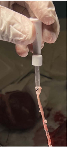
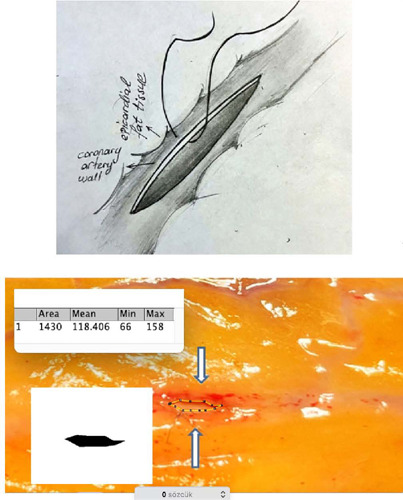
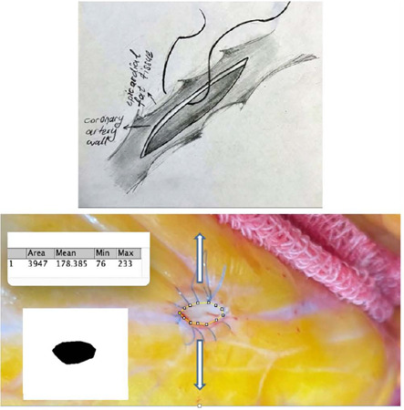
Methods: This study was conducted by performing two different fashions of anastomosis with a human saphenous vein graft on 24 various coronary segments of five postmortem porcine hearts. Each arteriotomy was used for both anastomotic techniques. In the first method, epicardial fat tissue around the coronary artery was involved to the saphenous vein anastomosis line (coronary wall and epicardial fat tissue [CWE] technique). In the second method, the saphenous vein graft was sutured to the coronary wall only, without involving epicardial fat tissue (only coronary wall [OCW] technique).The time it tookfor 30 cc of 0.9% isotonic saline solution to pass through the anastomosis in a free-flow fashion by gravity was measured following each technique. Additionally, the anastomotic areas in mm2 were measured and compared between the two techniques.
Results: The mean flow time for the CWE technique was 77.5 ± 21.4 seconds, whereas for the OCW technique, it was 87.2 ± 19.5 seconds (P<0.001). The flow rates were 23.2 ml/min and 20.6 ml/min, respectively. The anastomotic area was 3.947 mm2 for the CWE technique and 1.430 mm2 for the OCW technique.
Conclusion: When the sutures penetrate both the epicardial fat tissue and the coronary artery wall simultaneously, a larger anastomosis area can be created. Consequently, potentially better graft flow and hemodynamic performance could be achieved.

 求助内容:
求助内容: 应助结果提醒方式:


