Daniel Cromb, Tom Finck, Alexandra F Bonthrone, Alena Uus, Milou Van Poppel, Johannes Steinweg, David F Lloyd, Kuberan Pushparajah, Reza Razavi, Serena J Counsell, Mary Rutherford
{"title":"An exploratory fetal MRI study examining the impact of 22q11.2 microdeletion syndrome on early brain growth.","authors":"Daniel Cromb, Tom Finck, Alexandra F Bonthrone, Alena Uus, Milou Van Poppel, Johannes Steinweg, David F Lloyd, Kuberan Pushparajah, Reza Razavi, Serena J Counsell, Mary Rutherford","doi":"10.1186/s11689-025-09594-9","DOIUrl":null,"url":null,"abstract":"<p><strong>Background: </strong>Improved long-term outcomes, related to advances in surgical and clinical care of infants with congenital heart disease (CHD), has shifted focus onto the accompanying and later-onset cognitive and neuropsychiatric disorders in those who also have 22q11.2 deletion syndrome (22qDS). 22qDS is itself associated with neurodevelopmental impairments and altered brain growth. However, when brain growth in 22qDS first deviates from normal is unknown, and whether impaired brain development is primarily genetics-driven or a secondary consequence of the underlying CHD remains incompletely understood.</p><p><strong>Methods: </strong>In this small, exploratory study, we use fetal MRI to assess volumetric brain development in 22qDS by comparing fetal brain morphometry to a set of gestation and sex-matched healthy controls, and a cohort of gestation and sex-matched fetuses with the same CHD diagnoses but without 22q11.2 deletion. Structural T2-weighted fetal brain images were acquired using a 1.5T MRI scanner. MR scanner and sequence parameters were identical in all cohorts. Motion-corrected images underwent segmentation using an automated pipeline developed for fetal brain MRI. Total brain tissue volumes, volumes for four different tissue regions (cortical grey matter, white matter, deep grey matter and cerebellum), cerebrospinal fluid and total intracranial volumes were calculated.</p><p><strong>Results: </strong>Antenatal imaging was acquired between 29 and 35 weeks gestation. Thirty-three fetuses were included (7 22qDS; 14 isolated CHD; 12 healthy control). White matter volumes were significantly reduced in fetuses with 22qDS compared to control fetuses (p = 0.028), but not to those with CHD without 22q11.2 deletion (p = 0.09). Large effect-sizes were seen between the 22qDS and isolated CHD cohorts (D<sub>Cohen</sub> = 0.81), and between the 22qDS and control cohorts (D<sub>Cohen</sub> = 1.2) for white matter volumes. No significant differences were seen in volumes of other brain regions between groups.</p><p><strong>Conclusions: </strong>This exploratory study expands our existing knowledge on neurodevelopmental impairments in 22qDS to the fetal period by highlighting reduced white matter volumes compared to gestation and sex-matched control fetuses during this time-period. Our findings suggest that impaired white matter growth in fetuses with both 22qDS and CHD may not be fully explained by any underlying CHD.</p>","PeriodicalId":16530,"journal":{"name":"Journal of Neurodevelopmental Disorders","volume":"17 1","pages":"7"},"PeriodicalIF":4.0000,"publicationDate":"2025-02-12","publicationTypes":"Journal Article","fieldsOfStudy":null,"isOpenAccess":false,"openAccessPdf":"https://www.ncbi.nlm.nih.gov/pmc/articles/PMC11817260/pdf/","citationCount":"0","resultStr":null,"platform":"Semanticscholar","paperid":null,"PeriodicalName":"Journal of Neurodevelopmental Disorders","FirstCategoryId":"3","ListUrlMain":"https://doi.org/10.1186/s11689-025-09594-9","RegionNum":2,"RegionCategory":"医学","ArticlePicture":[],"TitleCN":null,"AbstractTextCN":null,"PMCID":null,"EPubDate":"","PubModel":"","JCR":"Q1","JCRName":"CLINICAL NEUROLOGY","Score":null,"Total":0}
引用次数: 0
Abstract
Background: Improved long-term outcomes, related to advances in surgical and clinical care of infants with congenital heart disease (CHD), has shifted focus onto the accompanying and later-onset cognitive and neuropsychiatric disorders in those who also have 22q11.2 deletion syndrome (22qDS). 22qDS is itself associated with neurodevelopmental impairments and altered brain growth. However, when brain growth in 22qDS first deviates from normal is unknown, and whether impaired brain development is primarily genetics-driven or a secondary consequence of the underlying CHD remains incompletely understood.
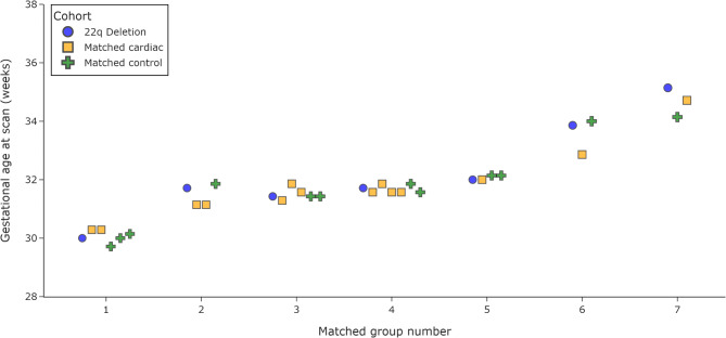
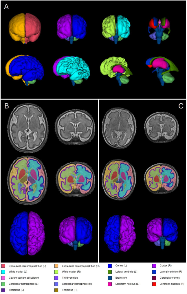
Methods: In this small, exploratory study, we use fetal MRI to assess volumetric brain development in 22qDS by comparing fetal brain morphometry to a set of gestation and sex-matched healthy controls, and a cohort of gestation and sex-matched fetuses with the same CHD diagnoses but without 22q11.2 deletion. Structural T2-weighted fetal brain images were acquired using a 1.5T MRI scanner. MR scanner and sequence parameters were identical in all cohorts. Motion-corrected images underwent segmentation using an automated pipeline developed for fetal brain MRI. Total brain tissue volumes, volumes for four different tissue regions (cortical grey matter, white matter, deep grey matter and cerebellum), cerebrospinal fluid and total intracranial volumes were calculated.
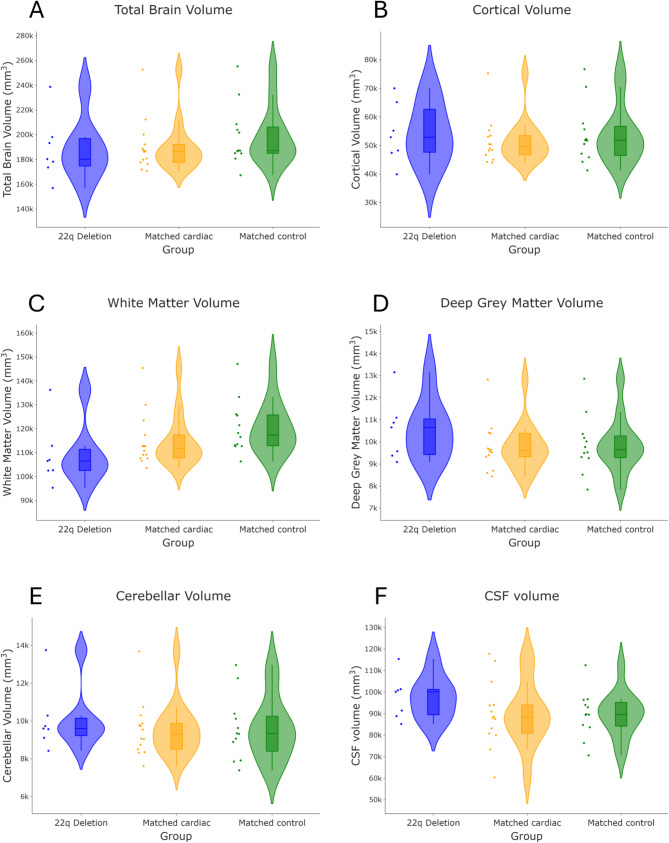
Results: Antenatal imaging was acquired between 29 and 35 weeks gestation. Thirty-three fetuses were included (7 22qDS; 14 isolated CHD; 12 healthy control). White matter volumes were significantly reduced in fetuses with 22qDS compared to control fetuses (p = 0.028), but not to those with CHD without 22q11.2 deletion (p = 0.09). Large effect-sizes were seen between the 22qDS and isolated CHD cohorts (DCohen = 0.81), and between the 22qDS and control cohorts (DCohen = 1.2) for white matter volumes. No significant differences were seen in volumes of other brain regions between groups.
Conclusions: This exploratory study expands our existing knowledge on neurodevelopmental impairments in 22qDS to the fetal period by highlighting reduced white matter volumes compared to gestation and sex-matched control fetuses during this time-period. Our findings suggest that impaired white matter growth in fetuses with both 22qDS and CHD may not be fully explained by any underlying CHD.
期刊介绍:
Journal of Neurodevelopmental Disorders is an open access journal that integrates current, cutting-edge research across a number of disciplines, including neurobiology, genetics, cognitive neuroscience, psychiatry and psychology. The journal’s primary focus is on the pathogenesis of neurodevelopmental disorders including autism, fragile X syndrome, tuberous sclerosis, Turner Syndrome, 22q Deletion Syndrome, Prader-Willi and Angelman Syndrome, Williams syndrome, lysosomal storage diseases, dyslexia, specific language impairment and fetal alcohol syndrome. With the discovery of specific genes underlying neurodevelopmental syndromes, the emergence of powerful tools for studying neural circuitry, and the development of new approaches for exploring molecular mechanisms, interdisciplinary research on the pathogenesis of neurodevelopmental disorders is now increasingly common. Journal of Neurodevelopmental Disorders provides a unique venue for researchers interested in comparing and contrasting mechanisms and characteristics related to the pathogenesis of the full range of neurodevelopmental disorders, sharpening our understanding of the etiology and relevant phenotypes of each condition.
 求助内容:
求助内容: 应助结果提醒方式:


