{"title":"The Impact of Contrast Media on Lumbar Spine Bone Mineral Density Measured by Quantitative Computed Tomography.","authors":"Lahari R Shetty, Kaushik Nayak, Priyanka","doi":"10.4314/ejhs.v34i5.3","DOIUrl":null,"url":null,"abstract":"<p><strong>Background: </strong>Osteoporosis is a bone disease caused by decrease in bone mineral density (BMD). Quantitative computed tomography (QCT) has proven to be an effective tool to measure the BMD of the lumbar spine. Therefore, the objective of the study is to investigate the impact of intravenous contrast media (CM) on BMD of lumbar spine measured by QCT.</p><p><strong>Methods: </strong>This is a prospective study and included a total of 141 patients (females: 71, males: 70) referred for contrast enhanced computed tomography (CECT) abdomen. First, the plain scan of abdomen was done. Contrast media was injected intravenously followed by acquisition of arterial and portovenous phase (PV) of abdomen. Plain, arterial and PV phases axial CT images were loaded on Philips BMD analysis application. A circular region of interest (ROI) measuring 30-40 mm<sup>2</sup> was placed at all five lumbar vertebrae (L1-L5) and value of BMD was obtained in mg/cm<sup>3</sup>.</p><p><strong>Results: </strong>Paired t-test was used to compare BMD in plain, arterial and PV phase. There was significant difference (p <0.05) in BMD (L1-L5) between plain (110.86±36.61 mg/cm<sup>3</sup>), arterial (117.04±37.95 mg/cm<sup>3</sup>) and PV phase (127.52±40.9 mg/cm<sup>3</sup>). The study also noted significant difference between males and females in BMD of lumbar spine (L1-L5) for plain and CECT abdomen (p <0.05).</p><p><strong>Conclusion: </strong>The BMD was highest for PV phase of the CECT abdomen. Therefore, the study concludes that BMD values are highly influenced by intravenous contrast media injections.</p>","PeriodicalId":12003,"journal":{"name":"Ethiopian Journal of Health Sciences","volume":"34 5","pages":"359-364"},"PeriodicalIF":1.5000,"publicationDate":"2024-09-01","publicationTypes":"Journal Article","fieldsOfStudy":null,"isOpenAccess":false,"openAccessPdf":"https://www.ncbi.nlm.nih.gov/pmc/articles/PMC11811390/pdf/","citationCount":"0","resultStr":null,"platform":"Semanticscholar","paperid":null,"PeriodicalName":"Ethiopian Journal of Health Sciences","FirstCategoryId":"1085","ListUrlMain":"https://doi.org/10.4314/ejhs.v34i5.3","RegionNum":0,"RegionCategory":null,"ArticlePicture":[],"TitleCN":null,"AbstractTextCN":null,"PMCID":null,"EPubDate":"","PubModel":"","JCR":"Q3","JCRName":"HEALTH CARE SCIENCES & SERVICES","Score":null,"Total":0}
引用次数: 0
Abstract
Background: Osteoporosis is a bone disease caused by decrease in bone mineral density (BMD). Quantitative computed tomography (QCT) has proven to be an effective tool to measure the BMD of the lumbar spine. Therefore, the objective of the study is to investigate the impact of intravenous contrast media (CM) on BMD of lumbar spine measured by QCT.
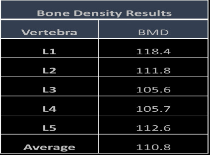
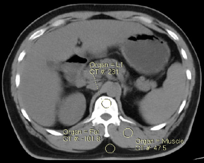
Methods: This is a prospective study and included a total of 141 patients (females: 71, males: 70) referred for contrast enhanced computed tomography (CECT) abdomen. First, the plain scan of abdomen was done. Contrast media was injected intravenously followed by acquisition of arterial and portovenous phase (PV) of abdomen. Plain, arterial and PV phases axial CT images were loaded on Philips BMD analysis application. A circular region of interest (ROI) measuring 30-40 mm2 was placed at all five lumbar vertebrae (L1-L5) and value of BMD was obtained in mg/cm3.
Results: Paired t-test was used to compare BMD in plain, arterial and PV phase. There was significant difference (p <0.05) in BMD (L1-L5) between plain (110.86±36.61 mg/cm3), arterial (117.04±37.95 mg/cm3) and PV phase (127.52±40.9 mg/cm3). The study also noted significant difference between males and females in BMD of lumbar spine (L1-L5) for plain and CECT abdomen (p <0.05).
Conclusion: The BMD was highest for PV phase of the CECT abdomen. Therefore, the study concludes that BMD values are highly influenced by intravenous contrast media injections.
期刊介绍:
Ethiopian Journal of Health Sciences is a general health science journal addressing clinical medicine, public health and biomedical sciences. Rarely, it covers veterinary medicine

 求助内容:
求助内容: 应助结果提醒方式:


