Chika Iguh, Julie Kim, Akudo Akaraonye, Amani Minja, Xin Qing
{"title":"An Unusual Case of Extracavitary/Solid Variant Primary Effusion Lymphoma With Associated Hemophagocytic Lymphohistiocytosis.","authors":"Chika Iguh, Julie Kim, Akudo Akaraonye, Amani Minja, Xin Qing","doi":"10.14740/jmc5084","DOIUrl":null,"url":null,"abstract":"<p><p>Primary effusion lymphoma (PEL) is a rare, aggressive large B-cell lymphoma variant that is invariably associated with human herpesvirus 8 (HHV8), predominantly in human immunodeficiency virus (HIV)-infected patients, and its oncogenicity is often augmented by coinfection with Epstein-Barr virus. It typically presents as a serous effusion in body cavities without detectable solid tumors. The extracavitary variant of PEL may represent a diagnostic challenge. A 37-year-old man with HIV/acquired immunodeficiency syndrome (AIDS) was transferred to our hospital for evaluation of a mediastinal mass with associated clinically diagnosed hemophagocytic lymphohistiocytosis (HLH), fever, pancytopenia, hepatosplenomegaly, retroperitoneal lymphadenopathy, and wasting syndrome. Contrast-enhanced computed tomography showed a large soft tissue mass extending along the middle/posterior mediastinum into the left hilum and a large left pleural effusion. Endoscopic fine-needle biopsy of the lesion showed sheets of large pleomorphic lymphoma cells with prominent nucleoli and abundant cytoplasm. These cells were also seen on the cytospin smear of pleural fluid. Immunohistochemical stains showed lymphoma cells positive for CD3 (small subset), CD45, CD138, MUM-1, and HHV8 and negative for CD5, CD20, CD30, ALK1, AE1/3, and PAX-5. The lymphoma cells were also positive for Epstein-Barr virus-encoded RNA (EBER) (<i>in situ</i> hybridization). Solid masses in extracavitary PEL have been shown to involve lymph nodes and/or solid organs such as the gastrointestinal tract, lung, liver, spleen, and skin, with a similar phenotype as classic PEL except that they may express B-cell markers with lower expression of CD45 and/or aberrant coexpression of T-cell antigens. This case illustrates the unusual manifestation of PEL as a mediastinal mass with associated HLH.</p>","PeriodicalId":101328,"journal":{"name":"Journal of medical cases","volume":"16 2","pages":"48-54"},"PeriodicalIF":0.9000,"publicationDate":"2025-02-01","publicationTypes":"Journal Article","fieldsOfStudy":null,"isOpenAccess":false,"openAccessPdf":"https://www.ncbi.nlm.nih.gov/pmc/articles/PMC11809604/pdf/","citationCount":"0","resultStr":null,"platform":"Semanticscholar","paperid":null,"PeriodicalName":"Journal of medical cases","FirstCategoryId":"1085","ListUrlMain":"https://doi.org/10.14740/jmc5084","RegionNum":0,"RegionCategory":null,"ArticlePicture":[],"TitleCN":null,"AbstractTextCN":null,"PMCID":null,"EPubDate":"2025/1/9 0:00:00","PubModel":"Epub","JCR":"","JCRName":"","Score":null,"Total":0}
引用次数: 0
Abstract
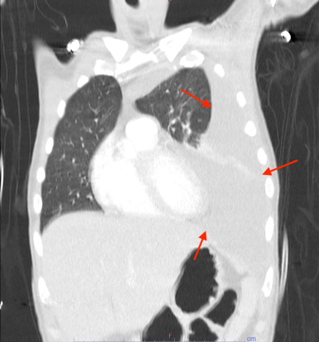
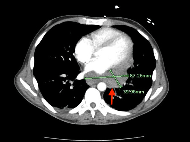
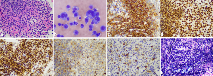
Primary effusion lymphoma (PEL) is a rare, aggressive large B-cell lymphoma variant that is invariably associated with human herpesvirus 8 (HHV8), predominantly in human immunodeficiency virus (HIV)-infected patients, and its oncogenicity is often augmented by coinfection with Epstein-Barr virus. It typically presents as a serous effusion in body cavities without detectable solid tumors. The extracavitary variant of PEL may represent a diagnostic challenge. A 37-year-old man with HIV/acquired immunodeficiency syndrome (AIDS) was transferred to our hospital for evaluation of a mediastinal mass with associated clinically diagnosed hemophagocytic lymphohistiocytosis (HLH), fever, pancytopenia, hepatosplenomegaly, retroperitoneal lymphadenopathy, and wasting syndrome. Contrast-enhanced computed tomography showed a large soft tissue mass extending along the middle/posterior mediastinum into the left hilum and a large left pleural effusion. Endoscopic fine-needle biopsy of the lesion showed sheets of large pleomorphic lymphoma cells with prominent nucleoli and abundant cytoplasm. These cells were also seen on the cytospin smear of pleural fluid. Immunohistochemical stains showed lymphoma cells positive for CD3 (small subset), CD45, CD138, MUM-1, and HHV8 and negative for CD5, CD20, CD30, ALK1, AE1/3, and PAX-5. The lymphoma cells were also positive for Epstein-Barr virus-encoded RNA (EBER) (in situ hybridization). Solid masses in extracavitary PEL have been shown to involve lymph nodes and/or solid organs such as the gastrointestinal tract, lung, liver, spleen, and skin, with a similar phenotype as classic PEL except that they may express B-cell markers with lower expression of CD45 and/or aberrant coexpression of T-cell antigens. This case illustrates the unusual manifestation of PEL as a mediastinal mass with associated HLH.

 求助内容:
求助内容: 应助结果提醒方式:


