{"title":"Radiographic Features of Spinal Meningioma and Schwannoma: A Novel Specific Feature-Ginkgo Leaf Sign.","authors":"Yu Toda, Masashi Miyazaki, Takaomi Kobayashi, Yoshiaki Egashira, Deokcheol Lee, Hideaki Hamanaka, Shigeo Ueda, Hiromu Yoshizato, Masatsugu Tsukamoto, Tomohito Yoshihara, Hirohito Hirata, Hiroaki Konishi, Tatsuya Tanaka, Koji Otani, Masaaki Mawatari, Tadatsugu Morimoto","doi":"10.22603/ssrr.2024-0059","DOIUrl":null,"url":null,"abstract":"<p><strong>Introduction: </strong>Meningiomas and schwannomas are common intradural-extramedullary spinal tumors. Because of their different origins, they necessitate different surgical procedures, which makes preoperative diagnosis important.</p><p><strong>Methods: </strong>In this study, clinical and imaging data for 62 patients diagnosed with either meningioma or schwannoma across multiple institutions were analyzed.</p><p><strong>Results: </strong>The average age of patients was older (67.6 vs. 58.9 years), and the frequency of females was higher (72% vs. 46%) for meningioma than for schwannoma. Meningiomas were mostly found in the thoracic region (84%), whereas schwannomas were commonly located in the lumbar region (54%). For each tumor type, specific radiological findings were identified. For meningiomas, findings included the ginkgo leaf sign (GLS) (sensitivity 58%, specificity 100%), oval shape (sensitivity 84%, specificity 63%), dural tail sign (DTS) (sensitivity 75%, specificity 100%), and intertumoral calcification (sensitivity 39%, specificity 100%). Combining GLS and DTS greatly improved sensitivity to 89% (specificity 100%). For schwannomas, specific findings included a lobule shape (sensitivity 25%, specificity 95%), dumbbell shape (sensitivity 54%, specificity 100%), and cystic changes (sensitivity 54%, specificity 97%).</p><p><strong>Conclusions: </strong>GLS may be a specific radiological feature for meningiomas and can aid in diagnosis when combined with DTS. Understanding these distinct radiological characteristics is valuable for preoperative differential diagnosis of intradural-extramedullary spinal tumors.</p>","PeriodicalId":22253,"journal":{"name":"Spine Surgery and Related Research","volume":"9 1","pages":"45-50"},"PeriodicalIF":1.2000,"publicationDate":"2024-07-10","publicationTypes":"Journal Article","fieldsOfStudy":null,"isOpenAccess":false,"openAccessPdf":"https://www.ncbi.nlm.nih.gov/pmc/articles/PMC11808241/pdf/","citationCount":"0","resultStr":null,"platform":"Semanticscholar","paperid":null,"PeriodicalName":"Spine Surgery and Related Research","FirstCategoryId":"1085","ListUrlMain":"https://doi.org/10.22603/ssrr.2024-0059","RegionNum":0,"RegionCategory":null,"ArticlePicture":[],"TitleCN":null,"AbstractTextCN":null,"PMCID":null,"EPubDate":"2025/1/27 0:00:00","PubModel":"eCollection","JCR":"Q3","JCRName":"SURGERY","Score":null,"Total":0}
引用次数: 0
Abstract
Introduction: Meningiomas and schwannomas are common intradural-extramedullary spinal tumors. Because of their different origins, they necessitate different surgical procedures, which makes preoperative diagnosis important.
Methods: In this study, clinical and imaging data for 62 patients diagnosed with either meningioma or schwannoma across multiple institutions were analyzed.
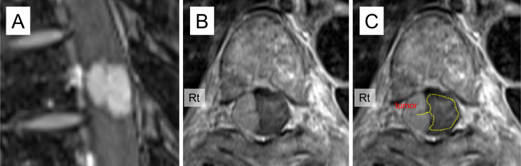
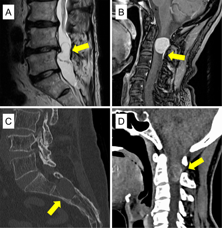
Results: The average age of patients was older (67.6 vs. 58.9 years), and the frequency of females was higher (72% vs. 46%) for meningioma than for schwannoma. Meningiomas were mostly found in the thoracic region (84%), whereas schwannomas were commonly located in the lumbar region (54%). For each tumor type, specific radiological findings were identified. For meningiomas, findings included the ginkgo leaf sign (GLS) (sensitivity 58%, specificity 100%), oval shape (sensitivity 84%, specificity 63%), dural tail sign (DTS) (sensitivity 75%, specificity 100%), and intertumoral calcification (sensitivity 39%, specificity 100%). Combining GLS and DTS greatly improved sensitivity to 89% (specificity 100%). For schwannomas, specific findings included a lobule shape (sensitivity 25%, specificity 95%), dumbbell shape (sensitivity 54%, specificity 100%), and cystic changes (sensitivity 54%, specificity 97%).
Conclusions: GLS may be a specific radiological feature for meningiomas and can aid in diagnosis when combined with DTS. Understanding these distinct radiological characteristics is valuable for preoperative differential diagnosis of intradural-extramedullary spinal tumors.

 求助内容:
求助内容: 应助结果提醒方式:


