{"title":"Difference of Disk Degeneration and Segmental Range of Motion due to Lumbar Disk Level among Age and Gender: 639 Asymptomatic Volunteer Data.","authors":"Tomohiro Yamada, Hiroaki Nakashima, Masaaki Machino, Yukihiro Matsuyama, Fumihiko Kato, Yasutsugu Yukawa","doi":"10.22603/ssrr.2024-0087","DOIUrl":null,"url":null,"abstract":"<p><strong>Introduction: </strong>There is limited evidence between lumbar disk degeneration and normal lumbar segmental range of motions (SRMs), because previous studies were skewed by age and lacked large cohort of asymptomatic data. We aimed to characterize the normal lumbar SRMs according to age and gender and determine its association with disk degeneration.</p><p><strong>Methods: </strong>A total of 639 healthy Japanese volunteers (≥50 individuals of each decade of age from 20 to 79) without any symptom or morphological spinal abnormalities, who underwent lumbar radiograph and magnetic resonance image (MRI), were selected retrospectively. SRMs were evaluated by the flexion-extension radiographs taken in the recumbent position. Disk degenerations were assessed according to the Pfirrmann grade using MRI T2 imaging.</p><p><strong>Results: </strong>The mean SRMs became larger in the lower lumbar level. The range of the mean SRMs was smallest at L1-2 and largest at L4-5: 6 to 9 degrees at L1/2, to peaking at 11-14 degrees at L4/5 in male, and 6-8 degrees at L1/2, to peaking at 11-17 degrees at L4/5 in female. Lumbar disk degeneration progressed faster with age in the lower lumbar spine than in the upper lumbar level. SRM did not change depending on the severity of disk degeneration in upper lumbar spine, but significantly decreased with progressive disk degeneration in the lower lumbar spine.</p><p><strong>Conclusions: </strong>These findings could help to identify the normal lumbar SRMs that might be useful to evaluate the instability or inflexibility in the clinical situation. Furthermore, our results demonstrated the transition of the normative lumbar SRMs based on age, gender, and lumbar level.</p>","PeriodicalId":22253,"journal":{"name":"Spine Surgery and Related Research","volume":"9 1","pages":"87-92"},"PeriodicalIF":1.2000,"publicationDate":"2024-07-10","publicationTypes":"Journal Article","fieldsOfStudy":null,"isOpenAccess":false,"openAccessPdf":"https://www.ncbi.nlm.nih.gov/pmc/articles/PMC11808234/pdf/","citationCount":"0","resultStr":null,"platform":"Semanticscholar","paperid":null,"PeriodicalName":"Spine Surgery and Related Research","FirstCategoryId":"1085","ListUrlMain":"https://doi.org/10.22603/ssrr.2024-0087","RegionNum":0,"RegionCategory":null,"ArticlePicture":[],"TitleCN":null,"AbstractTextCN":null,"PMCID":null,"EPubDate":"2025/1/27 0:00:00","PubModel":"eCollection","JCR":"Q3","JCRName":"SURGERY","Score":null,"Total":0}
引用次数: 0
Abstract
Introduction: There is limited evidence between lumbar disk degeneration and normal lumbar segmental range of motions (SRMs), because previous studies were skewed by age and lacked large cohort of asymptomatic data. We aimed to characterize the normal lumbar SRMs according to age and gender and determine its association with disk degeneration.
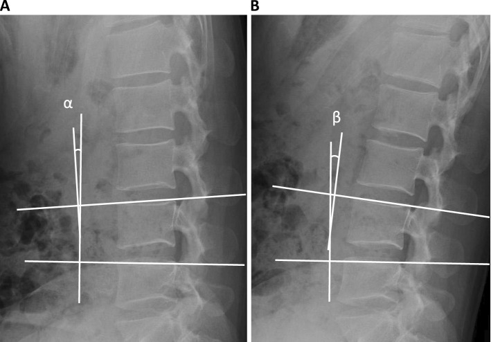
Methods: A total of 639 healthy Japanese volunteers (≥50 individuals of each decade of age from 20 to 79) without any symptom or morphological spinal abnormalities, who underwent lumbar radiograph and magnetic resonance image (MRI), were selected retrospectively. SRMs were evaluated by the flexion-extension radiographs taken in the recumbent position. Disk degenerations were assessed according to the Pfirrmann grade using MRI T2 imaging.
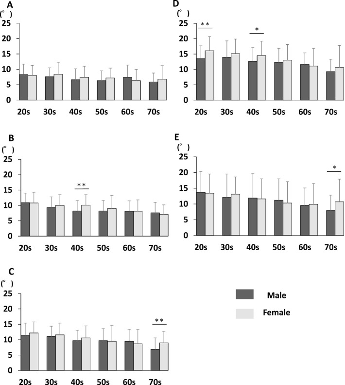
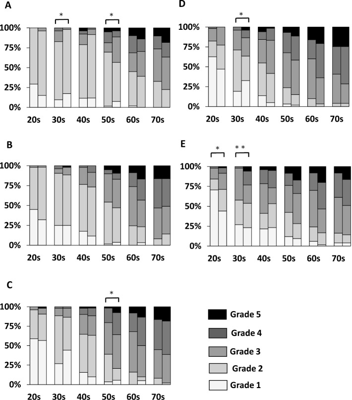
Results: The mean SRMs became larger in the lower lumbar level. The range of the mean SRMs was smallest at L1-2 and largest at L4-5: 6 to 9 degrees at L1/2, to peaking at 11-14 degrees at L4/5 in male, and 6-8 degrees at L1/2, to peaking at 11-17 degrees at L4/5 in female. Lumbar disk degeneration progressed faster with age in the lower lumbar spine than in the upper lumbar level. SRM did not change depending on the severity of disk degeneration in upper lumbar spine, but significantly decreased with progressive disk degeneration in the lower lumbar spine.
Conclusions: These findings could help to identify the normal lumbar SRMs that might be useful to evaluate the instability or inflexibility in the clinical situation. Furthermore, our results demonstrated the transition of the normative lumbar SRMs based on age, gender, and lumbar level.

 求助内容:
求助内容: 应助结果提醒方式:


