Variations in intracranial arterial anatomy of the circle of Willis and their association with arteriosclerosis in patients with ischemic cerebrovascular disease.
Bernhard P Berghout, Rüveyda F Soyupak, M Kamran Ikram, Daniel Bos
{"title":"Variations in intracranial arterial anatomy of the circle of Willis and their association with arteriosclerosis in patients with ischemic cerebrovascular disease.","authors":"Bernhard P Berghout, Rüveyda F Soyupak, M Kamran Ikram, Daniel Bos","doi":"10.1177/17474930251322678","DOIUrl":null,"url":null,"abstract":"<p><strong>Introduction: </strong>An estimated 20-31% of all people are born with a textbook anatomical configuration of the intracranial arteries comprising the Circle of Willis. Individuals with specific anatomical variants may be at elevated risk of intracranial arteriosclerosis, and possibly its sequelae of stroke and dementia, as the distribution of blood flow and pressure is known to be different in variants with missing arteries or arterial segments. Therefore, we studied the association of anatomical variation of intracranial arteries with arteriosclerosis.</p><p><strong>Methods: </strong>Between December 2005 and October 2010, 1126 patients (mean age: 62.3 (SD: ±14.0) years, 48.0% female) were recruited, 59.9% of whom had ischemic stroke and 40.1% a transient ischemic attack (TIA). Within the routine diagnostic work-up for stroke, patients underwent cranial computed tomography (CT) angiography. These images enabled a detailed visualization of intracranial arteries, which allowed for the assessment of the anatomical configuration of the cerebral arteries, the anterior and posterior communicating arteries, the internal carotids, and the vertebrobasilar arteries. In addition, these images facilitated the identification of intracranial arterial calcifications, the defining feature of intracranial arteriosclerosis. Binomial logistic regression models adjusting for age, sex, and ethnicity were constructed to assess associations between intracranial artery variations and presence of intracranial arterial calcifications.</p><p><strong>Results: </strong>An incomplete Circle of Willis, defined by aplasia of any arterial segment, was present in 875 (77.7%) patients. The most common variation found was aplasia of the right posterior communicating artery, in 52.0% of patients. Men more often presented with an incomplete anatomy as compared to women (adjusted odds ratio: 1.36 (95% CI = 1.02-1.81)). Intracranial artery calcification was present in 59.2% of patients. Incompleteness of the intracranial arteries was not associated with the presence of any intracranial artery calcification (0.95 (0.68-1.34)). However, specific variants were associated with specific locations of intracranial artery calcification: The prevalence of vertebrobasilar artery calcification was lower among those with fetal-type posterior cerebral artery compared to individuals with a normal posterior cerebral artery (0.61 (0.38-0.99)). The prevalence of vertebrobasilar artery calcification was higher among those with a-/hypoplasia of both posterior communicating arteries as compared to those with normal posterior communicating arteries (1.63 (1.00-2.66)). Furthermore, patients with a-/hypoplastic left A1-segments had a higher prevalence of right internal carotid artery calcification as compared to people with a normal left A1-segment (2.30 (1.00-5.26)).</p><p><strong>Conclusion: </strong>The prevalence of arteriosclerosis in the intracranial arteries on CT imaging varies among patients with certain anatomical variants of the intracranial arterial system. Specifically, arteriosclerosis in the right internal carotid artery and the vertebrobasilar arteries was more frequently observed in patients who had an a-/hypoplastic left anterior cerebral artery or a-/hypoplasia of both posterior communicating arteries, respectively. In addition, arteriosclerosis was less frequently observed among vertebrobasilar arteries of patients with a fetal-type posterior cerebral artery. Future longitudinal research is warranted regarding the anatomical configuration of intracranial arteries and the development of intracranial arteriosclerosis, as this line of research may reveal a novel group of people at elevated risk of cerebrovascular disease.</p>","PeriodicalId":14442,"journal":{"name":"International Journal of Stroke","volume":" ","pages":"843-851"},"PeriodicalIF":8.7000,"publicationDate":"2025-08-01","publicationTypes":"Journal Article","fieldsOfStudy":null,"isOpenAccess":false,"openAccessPdf":"https://www.ncbi.nlm.nih.gov/pmc/articles/PMC12264298/pdf/","citationCount":"0","resultStr":null,"platform":"Semanticscholar","paperid":null,"PeriodicalName":"International Journal of Stroke","FirstCategoryId":"3","ListUrlMain":"https://doi.org/10.1177/17474930251322678","RegionNum":2,"RegionCategory":"医学","ArticlePicture":[],"TitleCN":null,"AbstractTextCN":null,"PMCID":null,"EPubDate":"2025/2/27 0:00:00","PubModel":"Epub","JCR":"Q1","JCRName":"CLINICAL NEUROLOGY","Score":null,"Total":0}
引用次数: 0
Abstract
Introduction: An estimated 20-31% of all people are born with a textbook anatomical configuration of the intracranial arteries comprising the Circle of Willis. Individuals with specific anatomical variants may be at elevated risk of intracranial arteriosclerosis, and possibly its sequelae of stroke and dementia, as the distribution of blood flow and pressure is known to be different in variants with missing arteries or arterial segments. Therefore, we studied the association of anatomical variation of intracranial arteries with arteriosclerosis.
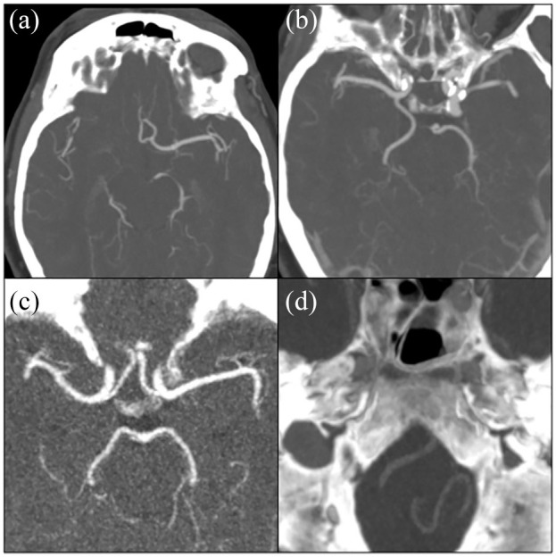
Methods: Between December 2005 and October 2010, 1126 patients (mean age: 62.3 (SD: ±14.0) years, 48.0% female) were recruited, 59.9% of whom had ischemic stroke and 40.1% a transient ischemic attack (TIA). Within the routine diagnostic work-up for stroke, patients underwent cranial computed tomography (CT) angiography. These images enabled a detailed visualization of intracranial arteries, which allowed for the assessment of the anatomical configuration of the cerebral arteries, the anterior and posterior communicating arteries, the internal carotids, and the vertebrobasilar arteries. In addition, these images facilitated the identification of intracranial arterial calcifications, the defining feature of intracranial arteriosclerosis. Binomial logistic regression models adjusting for age, sex, and ethnicity were constructed to assess associations between intracranial artery variations and presence of intracranial arterial calcifications.
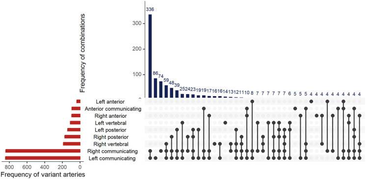
Results: An incomplete Circle of Willis, defined by aplasia of any arterial segment, was present in 875 (77.7%) patients. The most common variation found was aplasia of the right posterior communicating artery, in 52.0% of patients. Men more often presented with an incomplete anatomy as compared to women (adjusted odds ratio: 1.36 (95% CI = 1.02-1.81)). Intracranial artery calcification was present in 59.2% of patients. Incompleteness of the intracranial arteries was not associated with the presence of any intracranial artery calcification (0.95 (0.68-1.34)). However, specific variants were associated with specific locations of intracranial artery calcification: The prevalence of vertebrobasilar artery calcification was lower among those with fetal-type posterior cerebral artery compared to individuals with a normal posterior cerebral artery (0.61 (0.38-0.99)). The prevalence of vertebrobasilar artery calcification was higher among those with a-/hypoplasia of both posterior communicating arteries as compared to those with normal posterior communicating arteries (1.63 (1.00-2.66)). Furthermore, patients with a-/hypoplastic left A1-segments had a higher prevalence of right internal carotid artery calcification as compared to people with a normal left A1-segment (2.30 (1.00-5.26)).
Conclusion: The prevalence of arteriosclerosis in the intracranial arteries on CT imaging varies among patients with certain anatomical variants of the intracranial arterial system. Specifically, arteriosclerosis in the right internal carotid artery and the vertebrobasilar arteries was more frequently observed in patients who had an a-/hypoplastic left anterior cerebral artery or a-/hypoplasia of both posterior communicating arteries, respectively. In addition, arteriosclerosis was less frequently observed among vertebrobasilar arteries of patients with a fetal-type posterior cerebral artery. Future longitudinal research is warranted regarding the anatomical configuration of intracranial arteries and the development of intracranial arteriosclerosis, as this line of research may reveal a novel group of people at elevated risk of cerebrovascular disease.
期刊介绍:
The International Journal of Stroke is a welcome addition to the international stroke journal landscape in that it concentrates on the clinical aspects of stroke with basic science contributions in areas of clinical interest. Reviews of current topics are broadly based to encompass not only recent advances of global interest but also those which may be more important in certain regions and the journal regularly features items of news interest from all parts of the world. To facilitate the international nature of the journal, our Associate Editors from Europe, Asia, North America and South America coordinate segments of the journal.

 求助内容:
求助内容: 应助结果提醒方式:


