Blake A Gimbel, Jeffrey R Wozniak, Bryon A Mueller, Kent A Tuominen, Abigail M Ernst, Mary E Anthony, Erik de Water, Donovan J Roediger
{"title":"Regional hippocampal thinning and gyrification abnormalities and associated cognition in children with prenatal alcohol exposure.","authors":"Blake A Gimbel, Jeffrey R Wozniak, Bryon A Mueller, Kent A Tuominen, Abigail M Ernst, Mary E Anthony, Erik de Water, Donovan J Roediger","doi":"10.1186/s11689-025-09595-8","DOIUrl":null,"url":null,"abstract":"<p><strong>Background: </strong>Prenatal alcohol exposure (PAE) impacts hippocampal structure and function, contributing to deficits in memory and decision-making in affected individuals. Here, we evaluate hippocampal anomalies in children with PAE and an unexposed comparison group using advanced MRI methods that characterize hippocampal curvature and thickness.</p><p><strong>Methods: </strong>Participants, ages 8 to 16 years, included children with PAE (n = 48) and an unexposed comparison group (n = 46) who underwent a dysmorphology exam, neuropsychological assessment, and an MRI scan. Height, weight, head circumference, and dysmorphic facial features were evaluated. Of those with PAE, 4.2% had fetal alcohol syndrome (FAS), 22.9% had partial FAS, and 72.9% had alcohol-related neurodevelopmental disorder. Neuropsychological testing included measures of intelligence and memory functioning. T1-weighted anatomical data were processed with the Hippunfold pipeline, which \"unfolds\" the complex hippocampal structure onto a template surface and provides measures of thickness and gyrification/curvature at each vertex. Permutation Analysis of Linear Models (PALM) was used to test for group differences (PAE vs. comparison) in hippocampal thickness and gyrification at each vertex and also to assess correlations with cognitive functioning.</p><p><strong>Results: </strong>There were significant regional differences in thickness and gyrification across bilateral hippocampi, with the PAE group showing substantially thinner tissue and less curvature than the comparison group, especially in CA1 and subiculum regions. For those with PAE, thinner subicular tissue (bilateral) was associated with lower IQ. Also in the PAE group, lower episodic memory performance was associated with thinness in the right hippocampus, especially in the subiculum region. There were no significant regional hippocampal patterns that were associated with cognitive functioning for individuals in the unexposed comparison group.</p><p><strong>Conclusions: </strong>We used a novel MRI method to evaluate hippocampal structure in children with PAE and an unexposed comparison group. The data suggest that PAE disrupts hippocampal development, impacting both the early-stage folding of the structure and its ultimate thickness. The data also demonstrate that these developmental anomalies have functional consequences in terms of core memory functions as well as global intellectual functioning in children with PAE.</p>","PeriodicalId":16530,"journal":{"name":"Journal of Neurodevelopmental Disorders","volume":"17 1","pages":"5"},"PeriodicalIF":4.0000,"publicationDate":"2025-02-05","publicationTypes":"Journal Article","fieldsOfStudy":null,"isOpenAccess":false,"openAccessPdf":"https://www.ncbi.nlm.nih.gov/pmc/articles/PMC11796126/pdf/","citationCount":"0","resultStr":null,"platform":"Semanticscholar","paperid":null,"PeriodicalName":"Journal of Neurodevelopmental Disorders","FirstCategoryId":"3","ListUrlMain":"https://doi.org/10.1186/s11689-025-09595-8","RegionNum":2,"RegionCategory":"医学","ArticlePicture":[],"TitleCN":null,"AbstractTextCN":null,"PMCID":null,"EPubDate":"","PubModel":"","JCR":"Q1","JCRName":"CLINICAL NEUROLOGY","Score":null,"Total":0}
引用次数: 0
Abstract
Background: Prenatal alcohol exposure (PAE) impacts hippocampal structure and function, contributing to deficits in memory and decision-making in affected individuals. Here, we evaluate hippocampal anomalies in children with PAE and an unexposed comparison group using advanced MRI methods that characterize hippocampal curvature and thickness.
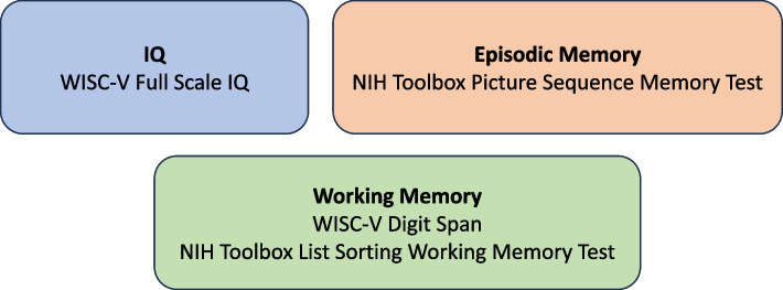
Methods: Participants, ages 8 to 16 years, included children with PAE (n = 48) and an unexposed comparison group (n = 46) who underwent a dysmorphology exam, neuropsychological assessment, and an MRI scan. Height, weight, head circumference, and dysmorphic facial features were evaluated. Of those with PAE, 4.2% had fetal alcohol syndrome (FAS), 22.9% had partial FAS, and 72.9% had alcohol-related neurodevelopmental disorder. Neuropsychological testing included measures of intelligence and memory functioning. T1-weighted anatomical data were processed with the Hippunfold pipeline, which "unfolds" the complex hippocampal structure onto a template surface and provides measures of thickness and gyrification/curvature at each vertex. Permutation Analysis of Linear Models (PALM) was used to test for group differences (PAE vs. comparison) in hippocampal thickness and gyrification at each vertex and also to assess correlations with cognitive functioning.
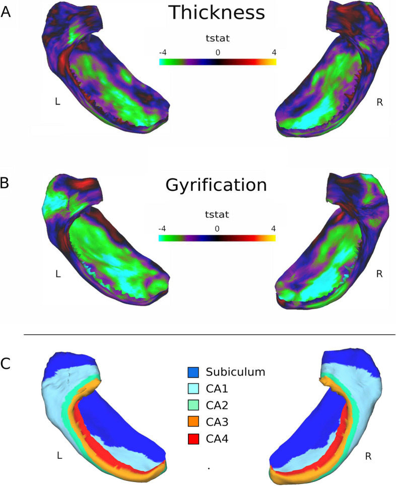
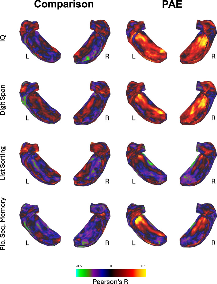
Results: There were significant regional differences in thickness and gyrification across bilateral hippocampi, with the PAE group showing substantially thinner tissue and less curvature than the comparison group, especially in CA1 and subiculum regions. For those with PAE, thinner subicular tissue (bilateral) was associated with lower IQ. Also in the PAE group, lower episodic memory performance was associated with thinness in the right hippocampus, especially in the subiculum region. There were no significant regional hippocampal patterns that were associated with cognitive functioning for individuals in the unexposed comparison group.
Conclusions: We used a novel MRI method to evaluate hippocampal structure in children with PAE and an unexposed comparison group. The data suggest that PAE disrupts hippocampal development, impacting both the early-stage folding of the structure and its ultimate thickness. The data also demonstrate that these developmental anomalies have functional consequences in terms of core memory functions as well as global intellectual functioning in children with PAE.
期刊介绍:
Journal of Neurodevelopmental Disorders is an open access journal that integrates current, cutting-edge research across a number of disciplines, including neurobiology, genetics, cognitive neuroscience, psychiatry and psychology. The journal’s primary focus is on the pathogenesis of neurodevelopmental disorders including autism, fragile X syndrome, tuberous sclerosis, Turner Syndrome, 22q Deletion Syndrome, Prader-Willi and Angelman Syndrome, Williams syndrome, lysosomal storage diseases, dyslexia, specific language impairment and fetal alcohol syndrome. With the discovery of specific genes underlying neurodevelopmental syndromes, the emergence of powerful tools for studying neural circuitry, and the development of new approaches for exploring molecular mechanisms, interdisciplinary research on the pathogenesis of neurodevelopmental disorders is now increasingly common. Journal of Neurodevelopmental Disorders provides a unique venue for researchers interested in comparing and contrasting mechanisms and characteristics related to the pathogenesis of the full range of neurodevelopmental disorders, sharpening our understanding of the etiology and relevant phenotypes of each condition.
 求助内容:
求助内容: 应助结果提醒方式:


