Sibel Demirel, Audrey Yan, Nicola Valsecchi, Jay Chhablani
{"title":"Imaging in Pachychoroid Disease","authors":"Sibel Demirel, Audrey Yan, Nicola Valsecchi, Jay Chhablani","doi":"10.4274/tjo.galenos.2024.40388","DOIUrl":null,"url":null,"abstract":"<p><p>The term pachychoroid was proposed as a term indicating an abnormal increase in choroidal thickness. Eyes presenting with pachychoroid changes often exhibit dilation of the large choroidal vessels, compressing the overlying choriocapillaris and Sattler’s layer. Pachychoroid spectrum diseases may present pathological findings such as pigment epitheliopathy, choroidal neovascularization (CNV), submacular serous detachment, and distinct choroidal and scleral alterations. Recent advancements in imaging modalities such as widefield indocyanine green angiography (WF-ICGA), optical coherence tomography angiography (OCTA), and enhanced depth imaging optical coherence tomography (OCT) have significantly improved our understanding of these conditions. WF-ICGA revealed venous outflow congestion in the peripheral retina as one of the characteristics of pachychoroid diseases. Scleral thickness measurements using ultrasound biomicroscopy and anterior segment OCT indicate that a thicker anterior sclera may contribute to choroidal congestion and disease pathogenesis. OCTA has emerged as a superior tool for identifying CNV and understanding the disease etiology, offering better sensitivity and specificity compared to traditional methods. These imaging advancements provide valuable insights into the structural and functional changes associated with pachychoroid diseases, potentially guiding future diagnostic and therapeutic strategies. The aim of the present review is to define the morphological characteristics of the pachychoroid spectrum of diseases, which share similar choroidal findings.</p>","PeriodicalId":23373,"journal":{"name":"Turkish Journal of Ophthalmology","volume":" ","pages":"36-48"},"PeriodicalIF":0.0000,"publicationDate":"2025-02-27","publicationTypes":"Journal Article","fieldsOfStudy":null,"isOpenAccess":false,"openAccessPdf":"https://www.ncbi.nlm.nih.gov/pmc/articles/PMC11866993/pdf/","citationCount":"0","resultStr":null,"platform":"Semanticscholar","paperid":null,"PeriodicalName":"Turkish Journal of Ophthalmology","FirstCategoryId":"1085","ListUrlMain":"https://doi.org/10.4274/tjo.galenos.2024.40388","RegionNum":0,"RegionCategory":null,"ArticlePicture":[],"TitleCN":null,"AbstractTextCN":null,"PMCID":null,"EPubDate":"2025/2/5 0:00:00","PubModel":"Epub","JCR":"Q3","JCRName":"Medicine","Score":null,"Total":0}
引用次数: 0
Abstract
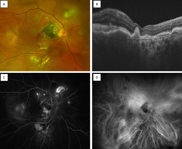
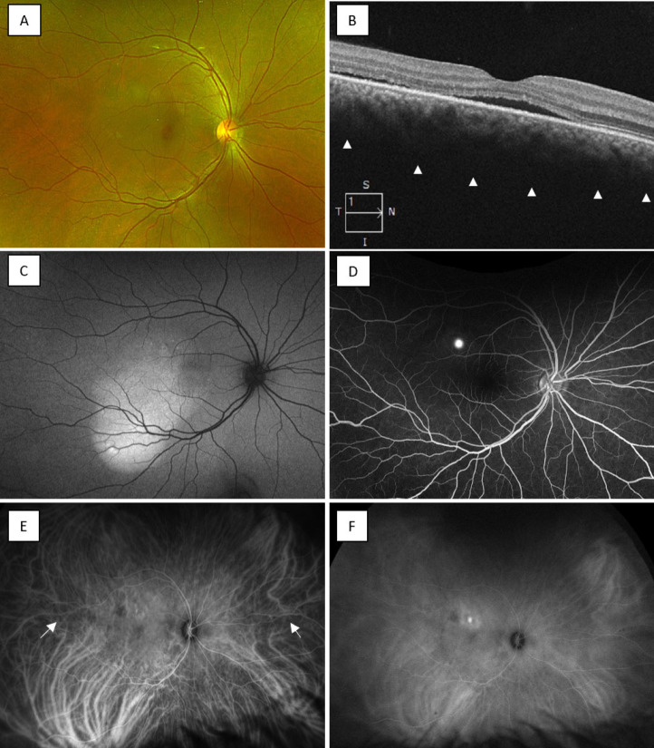
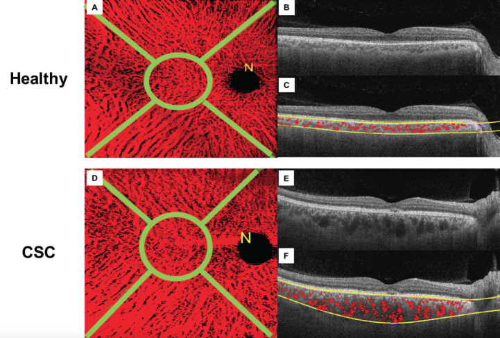
The term pachychoroid was proposed as a term indicating an abnormal increase in choroidal thickness. Eyes presenting with pachychoroid changes often exhibit dilation of the large choroidal vessels, compressing the overlying choriocapillaris and Sattler’s layer. Pachychoroid spectrum diseases may present pathological findings such as pigment epitheliopathy, choroidal neovascularization (CNV), submacular serous detachment, and distinct choroidal and scleral alterations. Recent advancements in imaging modalities such as widefield indocyanine green angiography (WF-ICGA), optical coherence tomography angiography (OCTA), and enhanced depth imaging optical coherence tomography (OCT) have significantly improved our understanding of these conditions. WF-ICGA revealed venous outflow congestion in the peripheral retina as one of the characteristics of pachychoroid diseases. Scleral thickness measurements using ultrasound biomicroscopy and anterior segment OCT indicate that a thicker anterior sclera may contribute to choroidal congestion and disease pathogenesis. OCTA has emerged as a superior tool for identifying CNV and understanding the disease etiology, offering better sensitivity and specificity compared to traditional methods. These imaging advancements provide valuable insights into the structural and functional changes associated with pachychoroid diseases, potentially guiding future diagnostic and therapeutic strategies. The aim of the present review is to define the morphological characteristics of the pachychoroid spectrum of diseases, which share similar choroidal findings.
期刊介绍:
The Turkish Journal of Ophthalmology (TJO) is the only scientific periodical publication of the Turkish Ophthalmological Association and has been published since January 1929. In its early years, the journal was published in Turkish and French. Although there were temporary interruptions in the publication of the journal due to various challenges, the Turkish Journal of Ophthalmology has been published continually from 1971 to the present. The target audience includes specialists and physicians in training in ophthalmology in all relevant disciplines.

 求助内容:
求助内容: 应助结果提醒方式:


