Retinal blood vessel segmentation and inpainting networks with multi-level self-attention
IF 4.9
2区 医学
Q1 ENGINEERING, BIOMEDICAL
引用次数: 0
Abstract
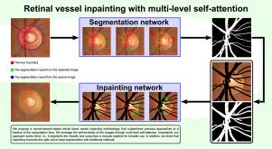
Improvement and restoration of retinal images are vital for clinical applications, from abnormality classification through segmentation to automated medical diagnosis. The major problem of retina restoration is estimating the image regions obscured by unwanted features, of which the most significant culprit is the blood vessel network. The challenge lies in the unavailability of true, unobstructed images. The commonly used methods apply masked filtering, dictionary-based approaches, or rely on an innate ability of machine learning models to deal with blood vessels. To solve the blind blood vessel inpainting problem, we propose a convolutional network architecture with multi-level self-attention capable of learning both segmentation and inpainting of blood vessels in retinal images. Furthermore, we introduce an efficient training method for the inpainting task with unknown ground truth. Our focus is on the optic nerve head region, which is essential in fundus analysis and glaucoma diagnosis. Our approach surpasses the state-of-the-art methods in blood vessel inpainting on the examined data while being trainable on personal computers. We examine the accuracy of vessel segmentation and the quality of inpainted images produced by our approach. The results show a statistically significant increase in segmentation accuracy of traditional methods after inpainting. In conclusion, we present a reliable vessel removal method applicable as a crucial first step in retinal segmentation, in the shape and color analysis of separated retinal vessels and background, in blood vessel detection, or in generating clear retinal background for generative methods.
多层次自关注视网膜血管分割与修复网络
从异常分类、分割到自动医学诊断,视网膜图像的改善和恢复对临床应用至关重要。视网膜修复的主要问题是如何估计被不需要的特征所遮挡的图像区域,其中最主要的罪魁祸首是血管网络。挑战在于无法获得真实的、无障碍的图像。常用的方法包括屏蔽过滤、基于字典的方法,或者依靠机器学习模型的固有能力来处理血管。为了解决血管的盲涂问题,我们提出了一种具有多层自关注的卷积网络结构,能够学习视网膜图像中血管的分割和涂漆。在此基础上,提出了一种有效的训练方法,用于未知真实值的喷漆任务。我们的重点是视神经头区域,这是必不可少的眼底分析和青光眼诊断。我们的方法超越了最先进的血管彩绘方法,同时可以在个人电脑上进行训练。我们检验了血管分割的准确性和我们的方法所产生的图像的质量。结果表明,与传统的分割方法相比,经过图像处理后的分割精度有了显著的提高。总之,我们提出了一种可靠的血管去除方法,适用于视网膜分割、分离的视网膜血管和背景的形状和颜色分析、血管检测或生成生成方法的清晰视网膜背景的关键第一步。
本文章由计算机程序翻译,如有差异,请以英文原文为准。
求助全文
约1分钟内获得全文
求助全文
来源期刊

Biomedical Signal Processing and Control
工程技术-工程:生物医学
CiteScore
9.80
自引率
13.70%
发文量
822
审稿时长
4 months
期刊介绍:
Biomedical Signal Processing and Control aims to provide a cross-disciplinary international forum for the interchange of information on research in the measurement and analysis of signals and images in clinical medicine and the biological sciences. Emphasis is placed on contributions dealing with the practical, applications-led research on the use of methods and devices in clinical diagnosis, patient monitoring and management.
Biomedical Signal Processing and Control reflects the main areas in which these methods are being used and developed at the interface of both engineering and clinical science. The scope of the journal is defined to include relevant review papers, technical notes, short communications and letters. Tutorial papers and special issues will also be published.
 求助内容:
求助内容: 应助结果提醒方式:


