{"title":"Automated Patient-specific Quality Assurance for Automated Segmentation of Organs at Risk in Nasopharyngeal Carcinoma Radiotherapy.","authors":"Yixuan Wang, Jiang Hu, Lixin Chen, Dandan Zhang, Jinhan Zhu","doi":"10.1177/10732748251318387","DOIUrl":null,"url":null,"abstract":"<p><strong>Introduction: </strong>Precision radiotherapy relies on accurate segmentation of tumor targets and organs at risk (OARs). Clinicians manually review automatically delineated structures on a case-by-case basis, a time-consuming process dependent on reviewer experience and alertness. This study proposes a general process for automated threshold generation for structural evaluation indicators and patient-specific quality assurance (QA) for automated segmentation of nasopharyngeal carcinoma (NPC).</p><p><strong>Methods: </strong>The patient-specific QA process for automated segmentation involves determining the confidence limit and error structure highlight stage. Three expert physicians segmented 17 OARs using computed tomography images of NPC and compared them using the Dice similarity coefficient, the maximum Hausdorff distance, and the mean distance to agreement. For each OAR, the 95% confidence interval was calculated as the confidence limit for each indicator. If two or more evaluation indicators (N2) or one or more evaluation indicators (N1) exceeded the confidence limits, the structure segmentation result was considered abnormal. The quantitative performances of these two methods were compared with those obtained by artificially introducing small/medium and serious errors.</p><p><strong>Results: </strong>The sensitivity, specificity, balanced accuracy, and F-score values for N2 were 0.944 ± 0.052, 0.827 ± 0.149, 0.886 ± 0.076, and 0.936 ± 0.045, respectively, whereas those for N1 were 0.955 ± 0.045, 0.788 ± 0.189, 0.878 ± 0.096, and 0.948 ± 0.035, respectively. N2 and N1 had small/medium error detection rates of 97.67 ± 0.04% and 98.67 ± 0.04%, respectively, with a serious error detection rate of 100%.</p><p><strong>Conclusion: </strong>The proposed automated patient-specific QA process effectively detected segmentation abnormalities, particularly serious errors. These are crucial for enhancing review efficiency and automated segmentation, and for improving physician confidence in automated segmentation.</p>","PeriodicalId":49093,"journal":{"name":"Cancer Control","volume":"32 ","pages":"10732748251318387"},"PeriodicalIF":2.6000,"publicationDate":"2025-01-01","publicationTypes":"Journal Article","fieldsOfStudy":null,"isOpenAccess":false,"openAccessPdf":"https://www.ncbi.nlm.nih.gov/pmc/articles/PMC11792024/pdf/","citationCount":"0","resultStr":null,"platform":"Semanticscholar","paperid":null,"PeriodicalName":"Cancer Control","FirstCategoryId":"3","ListUrlMain":"https://doi.org/10.1177/10732748251318387","RegionNum":4,"RegionCategory":"医学","ArticlePicture":[],"TitleCN":null,"AbstractTextCN":null,"PMCID":null,"EPubDate":"","PubModel":"","JCR":"Q3","JCRName":"ONCOLOGY","Score":null,"Total":0}
引用次数: 0
Abstract
Introduction: Precision radiotherapy relies on accurate segmentation of tumor targets and organs at risk (OARs). Clinicians manually review automatically delineated structures on a case-by-case basis, a time-consuming process dependent on reviewer experience and alertness. This study proposes a general process for automated threshold generation for structural evaluation indicators and patient-specific quality assurance (QA) for automated segmentation of nasopharyngeal carcinoma (NPC).
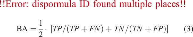
Methods: The patient-specific QA process for automated segmentation involves determining the confidence limit and error structure highlight stage. Three expert physicians segmented 17 OARs using computed tomography images of NPC and compared them using the Dice similarity coefficient, the maximum Hausdorff distance, and the mean distance to agreement. For each OAR, the 95% confidence interval was calculated as the confidence limit for each indicator. If two or more evaluation indicators (N2) or one or more evaluation indicators (N1) exceeded the confidence limits, the structure segmentation result was considered abnormal. The quantitative performances of these two methods were compared with those obtained by artificially introducing small/medium and serious errors.
Results: The sensitivity, specificity, balanced accuracy, and F-score values for N2 were 0.944 ± 0.052, 0.827 ± 0.149, 0.886 ± 0.076, and 0.936 ± 0.045, respectively, whereas those for N1 were 0.955 ± 0.045, 0.788 ± 0.189, 0.878 ± 0.096, and 0.948 ± 0.035, respectively. N2 and N1 had small/medium error detection rates of 97.67 ± 0.04% and 98.67 ± 0.04%, respectively, with a serious error detection rate of 100%.
Conclusion: The proposed automated patient-specific QA process effectively detected segmentation abnormalities, particularly serious errors. These are crucial for enhancing review efficiency and automated segmentation, and for improving physician confidence in automated segmentation.
期刊介绍:
Cancer Control is a JCR-ranked, peer-reviewed open access journal whose mission is to advance the prevention, detection, diagnosis, treatment, and palliative care of cancer by enabling researchers, doctors, policymakers, and other healthcare professionals to freely share research along the cancer control continuum. Our vision is a world where gold-standard cancer care is the norm, not the exception.


 求助内容:
求助内容: 应助结果提醒方式:


