SALL4 as a Useful Marker for the Distinction of Various Gestational Trophoblastic Disease Subtypes: Choriocarcinoma From Other Trophoblastic Lesions and Early Complete Hydatidiform Mole From Partial Mole and NonMolar Villi.
Alexis Trecourt, Marie Donzel, Lucie Gaillot-Durand, Pierre A Bolze, François Golfier, Pierre Descargues, Touria Hajri, Claire Mauduit, Mojgan Devouassoux-Shisheboran, Fabienne Allias
{"title":"SALL4 as a Useful Marker for the Distinction of Various Gestational Trophoblastic Disease Subtypes: Choriocarcinoma From Other Trophoblastic Lesions and Early Complete Hydatidiform Mole From Partial Mole and NonMolar Villi.","authors":"Alexis Trecourt, Marie Donzel, Lucie Gaillot-Durand, Pierre A Bolze, François Golfier, Pierre Descargues, Touria Hajri, Claire Mauduit, Mojgan Devouassoux-Shisheboran, Fabienne Allias","doi":"10.1097/PAS.0000000000002358","DOIUrl":null,"url":null,"abstract":"<p><p>The distinction between choriocarcinoma and residual trophoblastic cell proliferation from a complete hydatidiform mole/invasive mole (CHM/IM) without villi is challenging on curettage materials. We investigated whether SALL4 immunostaining could help differentiate various gestational trophoblastic diseases. Placental site nodules (PSN; n=10), atypical PSN (APSN; n=8), placental site trophoblastic tumors (PSTT; n=9), epithelioid trophoblastic tumors (ETT; n=5), gestational choriocarcinomas (n=31), partial hydatidiform moles (PHM; n=13), CHM/IM (n=47), and nonmolar products of conception (POC) (n=26) were included. SALL4 immunostaining was quantified (0 [1% to 10%], [11% to 100%]) and characterized (scattered single-cell or clustered nuclear positivity) in 2 locations: cytotrophoblast/intermediate trophoblast and villous stromal fibroblasts. A diffuse (11% to 100%) and clustered pattern of SALL4 immunostaining in cytotrophoblast/intermediate trophoblast was statistically associated with choriocarcinomas (74.2%, 23/31) as compared with PSN (0/10; P <0.0001), APSN (0/8; P =0.0002), PSTT (0/9; P <0.0001), ETT (0/5; P =0.0034), PHM (0/13; P <0.0001), CHM/IM (0/47; P <0.0001), and nonmolar POC (0/26; P <0.0001). Most nonchoriocarcinoma samples showed no SALL4 expression; when present, it was of low level (1% to 10%) and with a scattered single-cell staining in 3/9 PSTT (33%), 1/13 PHM (7.7%), 19/47 CHM/IM (40%), and 1/26 nonmolar POC (1.7%). These results were confirmed using a validation cohort. In addition, 66% (31/47) of CHM/IM villous stromal fibroblasts showed SALL4 expression (11% to 100%) (all before 14 gestational weeks), whereas this level of expression was never observed in PHM (0/13), nor in nonmolar POC (0/26; P <0.0001). Finally, a clustered and >10% SALL4 immunostaining in cytotrophoblast/intermediate trophoblast favors choriocarcinoma diagnosis. SALL4 expression in >10% villous stromal fibroblasts before 14 gestational weeks favors CHM/IM rather than PHM and nonmolar POC.</p>","PeriodicalId":7772,"journal":{"name":"American Journal of Surgical Pathology","volume":" ","pages":"417-428"},"PeriodicalIF":4.2000,"publicationDate":"2025-05-01","publicationTypes":"Journal Article","fieldsOfStudy":null,"isOpenAccess":false,"openAccessPdf":"https://www.ncbi.nlm.nih.gov/pmc/articles/PMC11984545/pdf/","citationCount":"0","resultStr":null,"platform":"Semanticscholar","paperid":null,"PeriodicalName":"American Journal of Surgical Pathology","FirstCategoryId":"3","ListUrlMain":"https://doi.org/10.1097/PAS.0000000000002358","RegionNum":1,"RegionCategory":"医学","ArticlePicture":[],"TitleCN":null,"AbstractTextCN":null,"PMCID":null,"EPubDate":"2025/1/29 0:00:00","PubModel":"Epub","JCR":"Q1","JCRName":"PATHOLOGY","Score":null,"Total":0}
引用次数: 0
Abstract
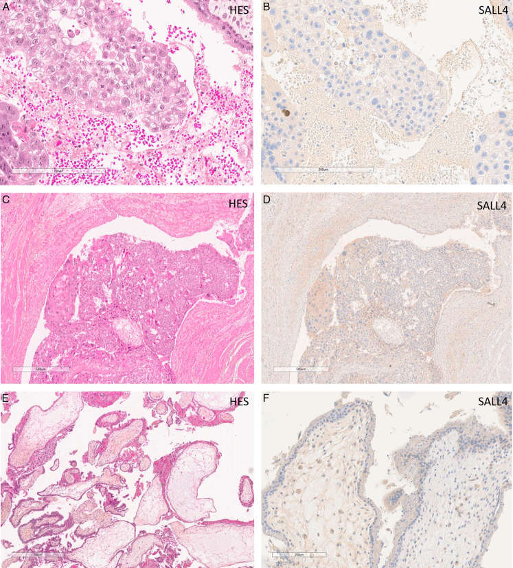
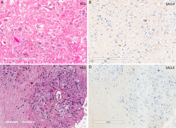
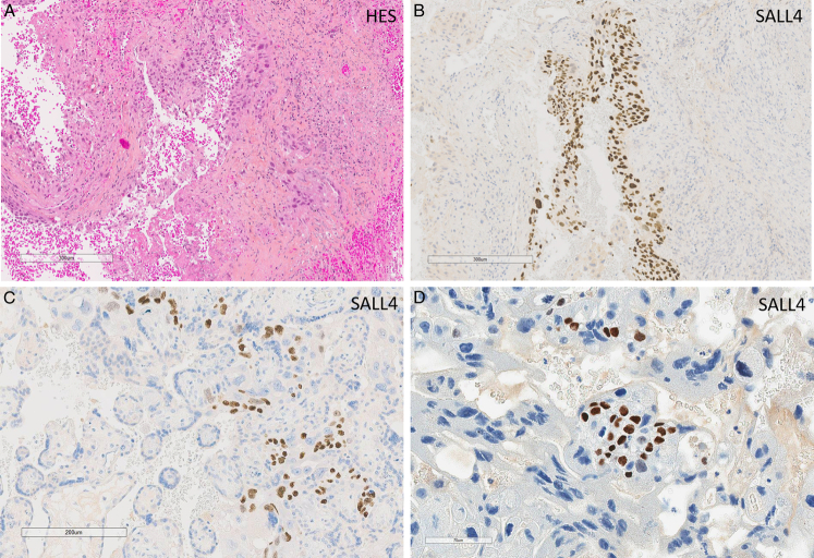
The distinction between choriocarcinoma and residual trophoblastic cell proliferation from a complete hydatidiform mole/invasive mole (CHM/IM) without villi is challenging on curettage materials. We investigated whether SALL4 immunostaining could help differentiate various gestational trophoblastic diseases. Placental site nodules (PSN; n=10), atypical PSN (APSN; n=8), placental site trophoblastic tumors (PSTT; n=9), epithelioid trophoblastic tumors (ETT; n=5), gestational choriocarcinomas (n=31), partial hydatidiform moles (PHM; n=13), CHM/IM (n=47), and nonmolar products of conception (POC) (n=26) were included. SALL4 immunostaining was quantified (0 [1% to 10%], [11% to 100%]) and characterized (scattered single-cell or clustered nuclear positivity) in 2 locations: cytotrophoblast/intermediate trophoblast and villous stromal fibroblasts. A diffuse (11% to 100%) and clustered pattern of SALL4 immunostaining in cytotrophoblast/intermediate trophoblast was statistically associated with choriocarcinomas (74.2%, 23/31) as compared with PSN (0/10; P <0.0001), APSN (0/8; P =0.0002), PSTT (0/9; P <0.0001), ETT (0/5; P =0.0034), PHM (0/13; P <0.0001), CHM/IM (0/47; P <0.0001), and nonmolar POC (0/26; P <0.0001). Most nonchoriocarcinoma samples showed no SALL4 expression; when present, it was of low level (1% to 10%) and with a scattered single-cell staining in 3/9 PSTT (33%), 1/13 PHM (7.7%), 19/47 CHM/IM (40%), and 1/26 nonmolar POC (1.7%). These results were confirmed using a validation cohort. In addition, 66% (31/47) of CHM/IM villous stromal fibroblasts showed SALL4 expression (11% to 100%) (all before 14 gestational weeks), whereas this level of expression was never observed in PHM (0/13), nor in nonmolar POC (0/26; P <0.0001). Finally, a clustered and >10% SALL4 immunostaining in cytotrophoblast/intermediate trophoblast favors choriocarcinoma diagnosis. SALL4 expression in >10% villous stromal fibroblasts before 14 gestational weeks favors CHM/IM rather than PHM and nonmolar POC.
期刊介绍:
The American Journal of Surgical Pathology has achieved worldwide recognition for its outstanding coverage of the state of the art in human surgical pathology. In each monthly issue, experts present original articles, review articles, detailed case reports, and special features, enhanced by superb illustrations. Coverage encompasses technical methods, diagnostic aids, and frozen-section diagnosis, in addition to detailed pathologic studies of a wide range of disease entities.
Official Journal of The Arthur Purdy Stout Society of Surgical Pathologists and The Gastrointestinal Pathology Society.

 求助内容:
求助内容: 应助结果提醒方式:


