Shabaz Sultan, Mark A J Gorris, Evgenia Martynova, Lieke L van der Woude, Franka Buytenhuijs, Sandra van Wilpe, Kiek Verrijp, Carl G Figdor, I Jolanda M de Vries, Johannes Textor
{"title":"ImmuNet: a segmentation-free machine learning pipeline for immune landscape phenotyping in tumors by multiplex imaging.","authors":"Shabaz Sultan, Mark A J Gorris, Evgenia Martynova, Lieke L van der Woude, Franka Buytenhuijs, Sandra van Wilpe, Kiek Verrijp, Carl G Figdor, I Jolanda M de Vries, Johannes Textor","doi":"10.1093/biomethods/bpae094","DOIUrl":null,"url":null,"abstract":"<p><p>Tissue specimens taken from primary tumors or metastases contain important information for diagnosis and treatment of cancer patients. Multiplex imaging allows <i>in situ</i> visualization of heterogeneous cell populations, such as immune cells, in tissue samples. Most image processing pipelines first segment cell boundaries and then measure marker expression to assign cell phenotypes. In dense tissue environments, this segmentation-first approach can be inaccurate due to segmentation errors or overlapping cells. Here, we introduce the machine-learning pipeline \"ImmuNet\", which identifies positions and phenotypes of cells without segmenting them. ImmuNet is easy to train: human annotators only need to click on an immune cell and score its expression of each marker-drawing a full cell outline is not required. We trained and evaluated ImmuNet on multiplex images from human tonsil, lung cancer, prostate cancer, melanoma, and bladder cancer tissue samples and found it to consistently achieve error rates below 5%-10% across tissue types, cell types, and tissue densities, outperforming a segmentation-based baseline method. Furthermore, we externally validate ImmuNet results by comparing them to flow cytometric cell count measurements from the same tissue. In summary, ImmuNet is an effective, simpler alternative to segmentation-based approaches when only cell positions and phenotypes, but not their shapes, are required for downstream analyses. Thus, ImmuNet helps researchers to analyze cell positions in multiplex tissue images more easily and accurately.</p>","PeriodicalId":36528,"journal":{"name":"Biology Methods and Protocols","volume":"10 1","pages":"bpae094"},"PeriodicalIF":1.3000,"publicationDate":"2024-12-20","publicationTypes":"Journal Article","fieldsOfStudy":null,"isOpenAccess":false,"openAccessPdf":"https://www.ncbi.nlm.nih.gov/pmc/articles/PMC11769680/pdf/","citationCount":"0","resultStr":null,"platform":"Semanticscholar","paperid":null,"PeriodicalName":"Biology Methods and Protocols","FirstCategoryId":"1085","ListUrlMain":"https://doi.org/10.1093/biomethods/bpae094","RegionNum":0,"RegionCategory":null,"ArticlePicture":[],"TitleCN":null,"AbstractTextCN":null,"PMCID":null,"EPubDate":"2025/1/1 0:00:00","PubModel":"eCollection","JCR":"Q3","JCRName":"BIOCHEMICAL RESEARCH METHODS","Score":null,"Total":0}
引用次数: 0
Abstract
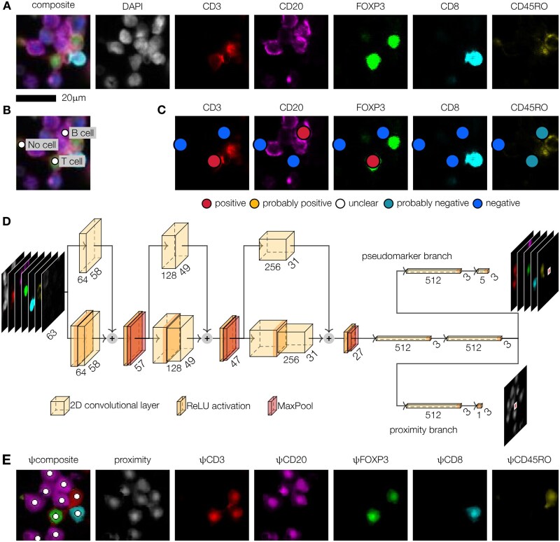
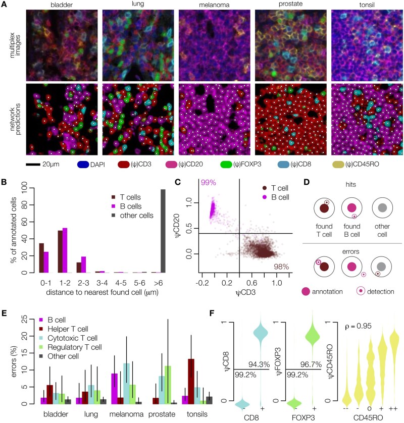
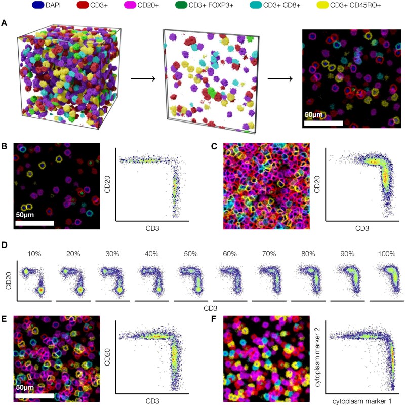
Tissue specimens taken from primary tumors or metastases contain important information for diagnosis and treatment of cancer patients. Multiplex imaging allows in situ visualization of heterogeneous cell populations, such as immune cells, in tissue samples. Most image processing pipelines first segment cell boundaries and then measure marker expression to assign cell phenotypes. In dense tissue environments, this segmentation-first approach can be inaccurate due to segmentation errors or overlapping cells. Here, we introduce the machine-learning pipeline "ImmuNet", which identifies positions and phenotypes of cells without segmenting them. ImmuNet is easy to train: human annotators only need to click on an immune cell and score its expression of each marker-drawing a full cell outline is not required. We trained and evaluated ImmuNet on multiplex images from human tonsil, lung cancer, prostate cancer, melanoma, and bladder cancer tissue samples and found it to consistently achieve error rates below 5%-10% across tissue types, cell types, and tissue densities, outperforming a segmentation-based baseline method. Furthermore, we externally validate ImmuNet results by comparing them to flow cytometric cell count measurements from the same tissue. In summary, ImmuNet is an effective, simpler alternative to segmentation-based approaches when only cell positions and phenotypes, but not their shapes, are required for downstream analyses. Thus, ImmuNet helps researchers to analyze cell positions in multiplex tissue images more easily and accurately.

 求助内容:
求助内容: 应助结果提醒方式:


