Enhanced Detection of Residual Breast Cancer Post-Excisional Biopsy: Comparative Analysis of Contrast-Enhanced MRI with and Without Diffusion-Weighted Imaging.
IF 2.2 4区 医学Q2 RADIOLOGY, NUCLEAR MEDICINE & MEDICAL IMAGING
Han Song Mun, Bong Joo Kang, Sung Hun Kim, Ga Eun Park
{"title":"Enhanced Detection of Residual Breast Cancer Post-Excisional Biopsy: Comparative Analysis of Contrast-Enhanced MRI with and Without Diffusion-Weighted Imaging.","authors":"Han Song Mun, Bong Joo Kang, Sung Hun Kim, Ga Eun Park","doi":"10.3390/tomography11010010","DOIUrl":null,"url":null,"abstract":"<p><strong>Objectives: </strong>To evaluate the effectiveness of breast MRI, including diffusion-weighted imaging (DWI), in detecting residual lesions in patients with malignancy after excisional biopsy.</p><p><strong>Methods: </strong>From January 2018 to December 2023, 3T breast MRI was performed to assess lesion morphology, residual size, and enhancement kinetics. The apparent diffusion coefficient (ADC) values were measured, and the diagnostic outcomes of CE-MRI, CE-MRI with DWI, mammography (MG), and ultrasound (US) were compared with clinical and histopathological data.</p><p><strong>Results: </strong>A total of 152 lesions were analyzed, with 36.2% showing residual malignancy. Both CE-MRI and CE-MRI with DWI effectively identified residual lesions, with significant differences in morphology, size, kinetic patterns, and ADC values (all <i>p</i> < 0.001). CE-MRI with DWI showed a sensitivity of 90.9% and an NPV of 93.6%, compared with 89.1% sensitivity and 92.2% NPV for CE-MRI alone. Sensitivities for MG and US were 57.1% and 38.7%, with NPVs of 64.7% and 59.6%, respectively. Diagnostic accuracy was highest for CE-MRI with DWI (80.9%), followed by CE-MRI (79.0%), MG (60.3%), and US (59.7%). The AUC for CE-MRI with DWI (0.831) was slightly higher than CE-MRI alone (0.811), though not significant (<i>p</i> = 0.095). AUCs for MG and US were lower at 0.623 and 0.563, with no significant difference between MG and US (<i>p</i> = 0.234).</p><p><strong>Conclusions: </strong>CE-MRI with DWI and CE-MRI alone were comparable and demonstrated excellent performance in discriminating between women with and without residual disease. Integrating CE-MRI with DWI could become a standard protocol for patients with suspected residual malignancy after excisional biopsy.</p>","PeriodicalId":51330,"journal":{"name":"Tomography","volume":"11 1","pages":""},"PeriodicalIF":2.2000,"publicationDate":"2025-01-20","publicationTypes":"Journal Article","fieldsOfStudy":null,"isOpenAccess":false,"openAccessPdf":"https://www.ncbi.nlm.nih.gov/pmc/articles/PMC11769435/pdf/","citationCount":"0","resultStr":null,"platform":"Semanticscholar","paperid":null,"PeriodicalName":"Tomography","FirstCategoryId":"3","ListUrlMain":"https://doi.org/10.3390/tomography11010010","RegionNum":4,"RegionCategory":"医学","ArticlePicture":[],"TitleCN":null,"AbstractTextCN":null,"PMCID":null,"EPubDate":"","PubModel":"","JCR":"Q2","JCRName":"RADIOLOGY, NUCLEAR MEDICINE & MEDICAL IMAGING","Score":null,"Total":0}
引用次数: 0
Abstract
Objectives: To evaluate the effectiveness of breast MRI, including diffusion-weighted imaging (DWI), in detecting residual lesions in patients with malignancy after excisional biopsy.
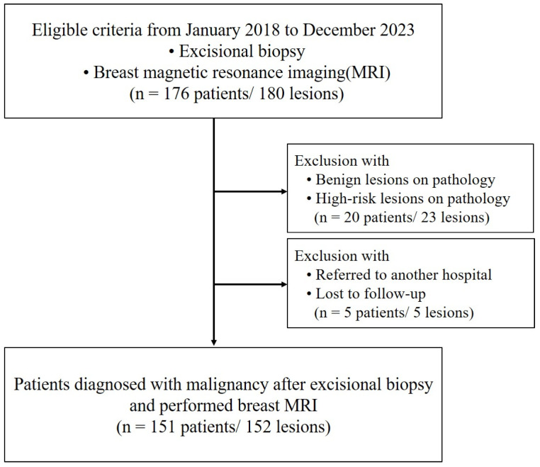
Methods: From January 2018 to December 2023, 3T breast MRI was performed to assess lesion morphology, residual size, and enhancement kinetics. The apparent diffusion coefficient (ADC) values were measured, and the diagnostic outcomes of CE-MRI, CE-MRI with DWI, mammography (MG), and ultrasound (US) were compared with clinical and histopathological data.
Results: A total of 152 lesions were analyzed, with 36.2% showing residual malignancy. Both CE-MRI and CE-MRI with DWI effectively identified residual lesions, with significant differences in morphology, size, kinetic patterns, and ADC values (all p < 0.001). CE-MRI with DWI showed a sensitivity of 90.9% and an NPV of 93.6%, compared with 89.1% sensitivity and 92.2% NPV for CE-MRI alone. Sensitivities for MG and US were 57.1% and 38.7%, with NPVs of 64.7% and 59.6%, respectively. Diagnostic accuracy was highest for CE-MRI with DWI (80.9%), followed by CE-MRI (79.0%), MG (60.3%), and US (59.7%). The AUC for CE-MRI with DWI (0.831) was slightly higher than CE-MRI alone (0.811), though not significant (p = 0.095). AUCs for MG and US were lower at 0.623 and 0.563, with no significant difference between MG and US (p = 0.234).
Conclusions: CE-MRI with DWI and CE-MRI alone were comparable and demonstrated excellent performance in discriminating between women with and without residual disease. Integrating CE-MRI with DWI could become a standard protocol for patients with suspected residual malignancy after excisional biopsy.
TomographyMedicine-Radiology, Nuclear Medicine and Imaging
CiteScore
2.70
自引率
10.50%
发文量
222
期刊介绍:
TomographyTM publishes basic (technical and pre-clinical) and clinical scientific articles which involve the advancement of imaging technologies. Tomography encompasses studies that use single or multiple imaging modalities including for example CT, US, PET, SPECT, MR and hyperpolarization technologies, as well as optical modalities (i.e. bioluminescence, photoacoustic, endomicroscopy, fiber optic imaging and optical computed tomography) in basic sciences, engineering, preclinical and clinical medicine.
Tomography also welcomes studies involving exploration and refinement of contrast mechanisms and image-derived metrics within and across modalities toward the development of novel imaging probes for image-based feedback and intervention. The use of imaging in biology and medicine provides unparalleled opportunities to noninvasively interrogate tissues to obtain real-time dynamic and quantitative information required for diagnosis and response to interventions and to follow evolving pathological conditions. As multi-modal studies and the complexities of imaging technologies themselves are ever increasing to provide advanced information to scientists and clinicians.
Tomography provides a unique publication venue allowing investigators the opportunity to more precisely communicate integrated findings related to the diverse and heterogeneous features associated with underlying anatomical, physiological, functional, metabolic and molecular genetic activities of normal and diseased tissue. Thus Tomography publishes peer-reviewed articles which involve the broad use of imaging of any tissue and disease type including both preclinical and clinical investigations. In addition, hardware/software along with chemical and molecular probe advances are welcome as they are deemed to significantly contribute towards the long-term goal of improving the overall impact of imaging on scientific and clinical discovery.

 求助内容:
求助内容: 应助结果提醒方式:


