Christophe Oosterbos, Ahmed M Radwan, Stefan Sunaert, Sophie Hoornaert, Anais Van Hoylandt, Robin Lemmens, Tom Theys
{"title":"Diffusion tensor imaging in peroneal neuropathy: a prospective, single-centre study.","authors":"Christophe Oosterbos, Ahmed M Radwan, Stefan Sunaert, Sophie Hoornaert, Anais Van Hoylandt, Robin Lemmens, Tom Theys","doi":"10.1136/bmjno-2024-000876","DOIUrl":null,"url":null,"abstract":"<p><strong>Objective: </strong>Diffusion tensor imaging (DTI) showed promising results in diagnosing upper limb neuropathies, but its value in patients with foot drop due to peroneal neuropathy has not yet been investigated. We aim to establish reference values for DTI metrics of the healthy peroneal nerve and to evaluate differences in DTI metrics between patients and healthy controls.</p><p><strong>Methods: </strong>Diffusion-weighted images (DWI) from 22 pathological nerves, 14 asymptomatic patients' nerves and 65 healthy peroneal nerves were processed for quantitative assessment of fractional anisotropy (FA), radial diffusivity (RD), axial diffusivity and mean diffusivity. Clinical baseline and follow-up data were prospectively collected for all patients.</p><p><strong>Results: </strong>Mean patient FA values (0.40, SD 0.08) were significantly lower compared with healthy controls (mean FA 0.44, SD 0.06). Mean patient RD values (0.98 10<sup>-3</sup> mm<sup>2</sup>/s, SD 0.21 10<sup>-3</sup> mm<sup>2</sup>/s) were significantly higher compared with healthy controls (mean RD 0.85 10<sup>-3</sup> mm<sup>2</sup>/s, SD 0.16 10<sup>-3</sup> mm<sup>2</sup>/s). FA values were significantly lower in patients with severe foot drop (mean FA 0.40, SD 0.06) compared with non-severe foot drop (mean FA 0.48, SD 0.05).</p><p><strong>Conclusion: </strong>Based on these results, DTI appears to aid in the differential diagnostic process of patients with peroneal neuropathy. Future studies should focus on automation of DWI processing, confirm the results in larger patient groups and try to establish reliable cut-off values for DTI metrics.</p>","PeriodicalId":52754,"journal":{"name":"BMJ Neurology Open","volume":"7 1","pages":"e000876"},"PeriodicalIF":2.4000,"publicationDate":"2025-01-09","publicationTypes":"Journal Article","fieldsOfStudy":null,"isOpenAccess":false,"openAccessPdf":"https://www.ncbi.nlm.nih.gov/pmc/articles/PMC11751924/pdf/","citationCount":"0","resultStr":null,"platform":"Semanticscholar","paperid":null,"PeriodicalName":"BMJ Neurology Open","FirstCategoryId":"1085","ListUrlMain":"https://doi.org/10.1136/bmjno-2024-000876","RegionNum":0,"RegionCategory":null,"ArticlePicture":[],"TitleCN":null,"AbstractTextCN":null,"PMCID":null,"EPubDate":"2025/1/1 0:00:00","PubModel":"eCollection","JCR":"Q3","JCRName":"CLINICAL NEUROLOGY","Score":null,"Total":0}
引用次数: 0
Abstract
Objective: Diffusion tensor imaging (DTI) showed promising results in diagnosing upper limb neuropathies, but its value in patients with foot drop due to peroneal neuropathy has not yet been investigated. We aim to establish reference values for DTI metrics of the healthy peroneal nerve and to evaluate differences in DTI metrics between patients and healthy controls.
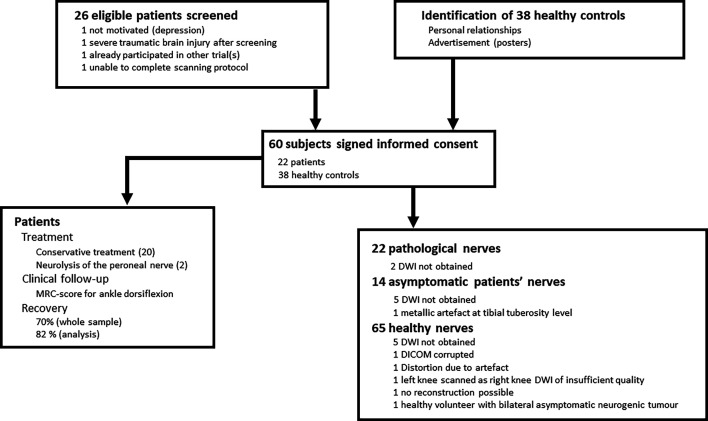
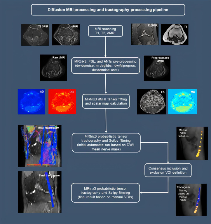
Methods: Diffusion-weighted images (DWI) from 22 pathological nerves, 14 asymptomatic patients' nerves and 65 healthy peroneal nerves were processed for quantitative assessment of fractional anisotropy (FA), radial diffusivity (RD), axial diffusivity and mean diffusivity. Clinical baseline and follow-up data were prospectively collected for all patients.
Results: Mean patient FA values (0.40, SD 0.08) were significantly lower compared with healthy controls (mean FA 0.44, SD 0.06). Mean patient RD values (0.98 10-3 mm2/s, SD 0.21 10-3 mm2/s) were significantly higher compared with healthy controls (mean RD 0.85 10-3 mm2/s, SD 0.16 10-3 mm2/s). FA values were significantly lower in patients with severe foot drop (mean FA 0.40, SD 0.06) compared with non-severe foot drop (mean FA 0.48, SD 0.05).
Conclusion: Based on these results, DTI appears to aid in the differential diagnostic process of patients with peroneal neuropathy. Future studies should focus on automation of DWI processing, confirm the results in larger patient groups and try to establish reliable cut-off values for DTI metrics.

 求助内容:
求助内容: 应助结果提醒方式:


