{"title":"CHROMOPHOBE HEPATOCELLULAR CARCINOMA: DIAGNOSTIC CHALLENGES.","authors":"Sana Ben-Slama, Ines Mallek, Eya Ghorbeli, Mohamed Hajri, Taher Labidi, Hafedh Mestiri, Ahlem Lahmar, Dhouha Bacha","doi":"10.1590/0102-6720202400069e1863","DOIUrl":null,"url":null,"abstract":"<p><strong>Background: </strong>Hepatocellular carcinoma (HCC) encompasses rare variants like chromophobe hepatocellular carcinoma (CHCC) characterized by distinct histological features and molecular profiles.</p><p><strong>Case report: </strong>A 56-year-old male with chronic hepatitis C, presenting pain in the right hypochondrium. Imaging revealed a solitary liver lesion, subsequently resected and histologically diagnosed as HCC. Macroscopic examination found a 4×4 cm encapsulated liver nodule with necrotic areas, surrounded by numerous smaller satellite nodules in Segment 6. The liver was in micronodular cirrhosis. Histologically, the tumor had focal trabecular or pseudoglandular patterns within a vascularized stroma. The cells were large, with clear to eosinophilic cytoplasm and hyperchromatic and pleomorphic nuclei with focal anaplastic features. No vascular invasion was noted in adjacent cirrhotic liver tissue.</p><p><strong>Results: </strong>The final diagnosis was CHCC. Due to its rarity and overlapping characteristics with other hepatic tumors, CHCC poses diagnostic challenges. Accurate diagnosis necessitates thorough histopathological assessment and molecular testing. The identification of the alternative lengthening of telomeres phenotype may distinguish CHCC from conventional HCC and hold potential implications for targeted therapeutic approaches.</p><p><strong>Conclusions: </strong>Recognition of HCC variants is critical for effective management and underscores the need for continued research into its clinical behavior and therapeutic responses.</p>","PeriodicalId":72298,"journal":{"name":"Arquivos brasileiros de cirurgia digestiva : ABCD = Brazilian archives of digestive surgery","volume":"37 ","pages":"e1863"},"PeriodicalIF":1.8000,"publicationDate":"2025-01-20","publicationTypes":"Journal Article","fieldsOfStudy":null,"isOpenAccess":false,"openAccessPdf":"https://www.ncbi.nlm.nih.gov/pmc/articles/PMC11745479/pdf/","citationCount":"0","resultStr":null,"platform":"Semanticscholar","paperid":null,"PeriodicalName":"Arquivos brasileiros de cirurgia digestiva : ABCD = Brazilian archives of digestive surgery","FirstCategoryId":"1085","ListUrlMain":"https://doi.org/10.1590/0102-6720202400069e1863","RegionNum":0,"RegionCategory":null,"ArticlePicture":[],"TitleCN":null,"AbstractTextCN":null,"PMCID":null,"EPubDate":"2025/1/1 0:00:00","PubModel":"eCollection","JCR":"","JCRName":"","Score":null,"Total":0}
引用次数: 0
Abstract
Background: Hepatocellular carcinoma (HCC) encompasses rare variants like chromophobe hepatocellular carcinoma (CHCC) characterized by distinct histological features and molecular profiles.
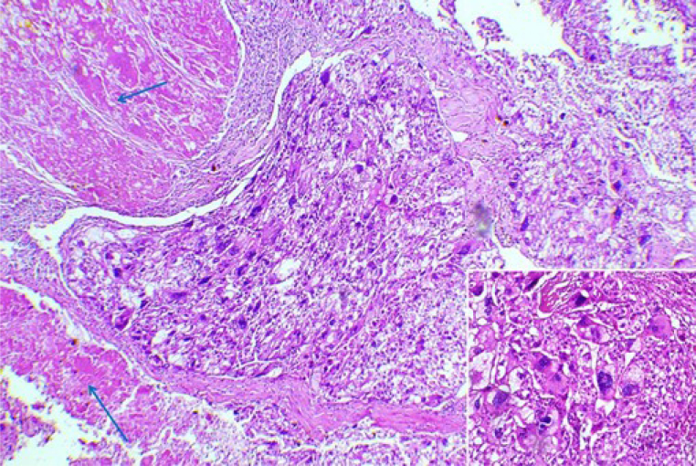
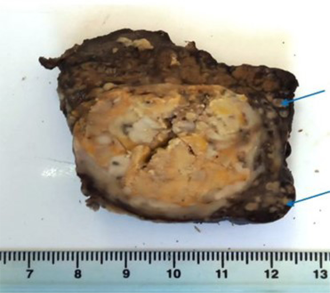
Case report: A 56-year-old male with chronic hepatitis C, presenting pain in the right hypochondrium. Imaging revealed a solitary liver lesion, subsequently resected and histologically diagnosed as HCC. Macroscopic examination found a 4×4 cm encapsulated liver nodule with necrotic areas, surrounded by numerous smaller satellite nodules in Segment 6. The liver was in micronodular cirrhosis. Histologically, the tumor had focal trabecular or pseudoglandular patterns within a vascularized stroma. The cells were large, with clear to eosinophilic cytoplasm and hyperchromatic and pleomorphic nuclei with focal anaplastic features. No vascular invasion was noted in adjacent cirrhotic liver tissue.
Results: The final diagnosis was CHCC. Due to its rarity and overlapping characteristics with other hepatic tumors, CHCC poses diagnostic challenges. Accurate diagnosis necessitates thorough histopathological assessment and molecular testing. The identification of the alternative lengthening of telomeres phenotype may distinguish CHCC from conventional HCC and hold potential implications for targeted therapeutic approaches.
Conclusions: Recognition of HCC variants is critical for effective management and underscores the need for continued research into its clinical behavior and therapeutic responses.


 求助内容:
求助内容: 应助结果提醒方式:


