Adam Dobek, Mateusz Kobierecki, Patryk Wieczorek, Oliwia Grząsiak, Wojciech Ciesielski, Adam Fabisiak, Ludomir Stefańczyk
{"title":"Contrast-enhanced ultrasonography as a method of monitoring focal liver lesions - initial report.","authors":"Adam Dobek, Mateusz Kobierecki, Patryk Wieczorek, Oliwia Grząsiak, Wojciech Ciesielski, Adam Fabisiak, Ludomir Stefańczyk","doi":"10.5114/ceh.2024.140449","DOIUrl":null,"url":null,"abstract":"<p><strong>Aim of the study: </strong>Hepatocellular adenoma (HCA) and focal nodular hyperplasia (FNH) are benign liver tumors. Hepatocellular adenoma has potential for growth, metaplasia and rupture; therefore, it should be monitored long term. In the current guidelines biopsy is not recommended in the standard diagnostic protocol. Magnetic resonance imaging (MRI) is accepted as standard in diagnostics and monitoring of these lesions. The aim of the study was to compare contrast-enhanced ultrasound (CEUS) and MRI in imaging of these tumors and determine whether CEUS can be useful in monitoring benign liver tumors.</p><p><strong>Material and methods: </strong>A retrospective analysis of 47 patients with HCA (32 tumors) and FNH (27 tumors) was carried out. A comparison between MRI and CEUS in predicting malignant transformation was performed.</p><p><strong>Results: </strong>A similar tumor enhancement profile to unchanged liver parenchyma was observed in both groups. The difference in the arterial phase was on average up to 30 dB. After 20-30 s, the enhancement of HCA and FNH in relation to the liver parenchyma was similar (difference up to 4-5 dB). Homogeneity and equalization of the tumor to background enhancement was observed until the end of the examination. The discriminative feature is the presence of a non-contrasting central fibrous scar observed in both imaging methods in the FNH group.</p><p><strong>Conclusions: </strong>CEUS can be a promising method in monitoring focal liver lesions due to low cost and low risk of complications. It is essential to analyze the early arterial phase up to 30 s to demonstrate homogeneous enhancement of the tumor and potential presence of a wash-out effect during later phases of examination.</p>","PeriodicalId":10281,"journal":{"name":"Clinical and Experimental Hepatology","volume":"10 2","pages":"120-128"},"PeriodicalIF":1.7000,"publicationDate":"2024-03-01","publicationTypes":"Journal Article","fieldsOfStudy":null,"isOpenAccess":false,"openAccessPdf":"https://www.ncbi.nlm.nih.gov/pmc/articles/PMC11748225/pdf/","citationCount":"0","resultStr":null,"platform":"Semanticscholar","paperid":null,"PeriodicalName":"Clinical and Experimental Hepatology","FirstCategoryId":"1085","ListUrlMain":"https://doi.org/10.5114/ceh.2024.140449","RegionNum":0,"RegionCategory":null,"ArticlePicture":[],"TitleCN":null,"AbstractTextCN":null,"PMCID":null,"EPubDate":"2024/6/11 0:00:00","PubModel":"Epub","JCR":"Q3","JCRName":"GASTROENTEROLOGY & HEPATOLOGY","Score":null,"Total":0}
引用次数: 0
Abstract
Aim of the study: Hepatocellular adenoma (HCA) and focal nodular hyperplasia (FNH) are benign liver tumors. Hepatocellular adenoma has potential for growth, metaplasia and rupture; therefore, it should be monitored long term. In the current guidelines biopsy is not recommended in the standard diagnostic protocol. Magnetic resonance imaging (MRI) is accepted as standard in diagnostics and monitoring of these lesions. The aim of the study was to compare contrast-enhanced ultrasound (CEUS) and MRI in imaging of these tumors and determine whether CEUS can be useful in monitoring benign liver tumors.
Material and methods: A retrospective analysis of 47 patients with HCA (32 tumors) and FNH (27 tumors) was carried out. A comparison between MRI and CEUS in predicting malignant transformation was performed.
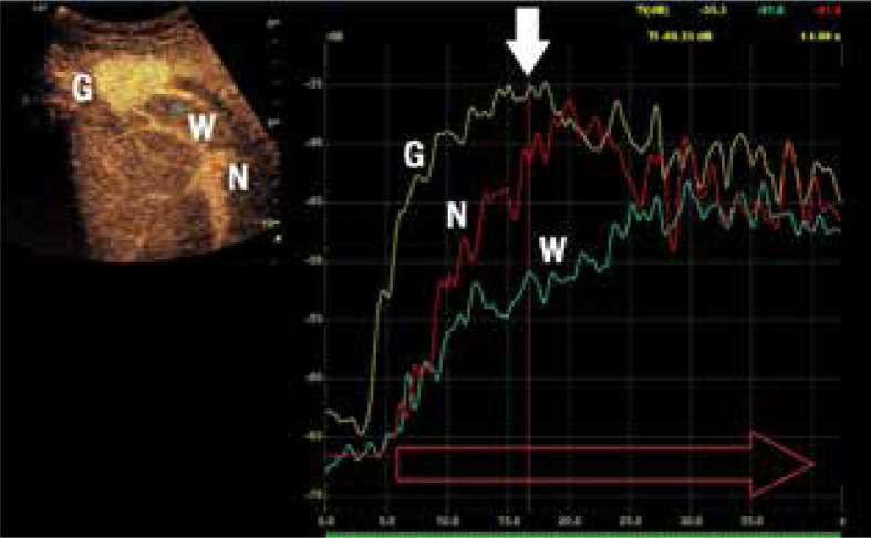
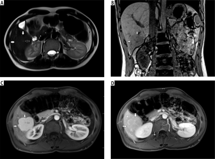
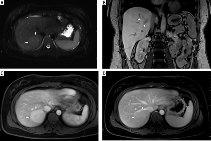
Results: A similar tumor enhancement profile to unchanged liver parenchyma was observed in both groups. The difference in the arterial phase was on average up to 30 dB. After 20-30 s, the enhancement of HCA and FNH in relation to the liver parenchyma was similar (difference up to 4-5 dB). Homogeneity and equalization of the tumor to background enhancement was observed until the end of the examination. The discriminative feature is the presence of a non-contrasting central fibrous scar observed in both imaging methods in the FNH group.
Conclusions: CEUS can be a promising method in monitoring focal liver lesions due to low cost and low risk of complications. It is essential to analyze the early arterial phase up to 30 s to demonstrate homogeneous enhancement of the tumor and potential presence of a wash-out effect during later phases of examination.
期刊介绍:
Clinical and Experimental Hepatology – quarterly of the Polish Association for Study of Liver – is a scientific and educational, peer-reviewed journal publishing original and review papers describing clinical and basic investigations in the field of hepatology.

 求助内容:
求助内容: 应助结果提醒方式:


