Pietro Fusari, Federico Guerri, Matteo Arcari, Andrea Sardella
{"title":"A rare case of bone lesion: Mandible's fibrous dysplasia.","authors":"Pietro Fusari, Federico Guerri, Matteo Arcari, Andrea Sardella","doi":"10.4103/njms.njms_35_23","DOIUrl":null,"url":null,"abstract":"<p><p>Fibrous dysplasia is a rare genetic syndrome that affects bone tissue. This pathology replaces the mineralized matrix of the bone affected with connective and fibrous tissue. This article describes a mandibular fibrous osseous dysplasia case and its surgical treatment. A 45-year-old woman complained about a slow development of swelling of the left mandibular bone. The orthopantomography (OPT) and the cone beam computed tomography (CBCT) revealed a well-circumscribed sclerotic lesion with a ground-glass appearance apical to the 3.5 element. The surgery was performed to excise the lesion. Anatomopathological examination of tissue confirmed the suspects among the diagnosis of fibrous dysplasia. The patient underwent to follow-up of 4 years, and no recurrences were found. In the absence of a univocal consensus on therapy, surgery remains the treatment of choice for unifocal forms.</p>","PeriodicalId":101444,"journal":{"name":"National journal of maxillofacial surgery","volume":"15 3","pages":"518-520"},"PeriodicalIF":0.0000,"publicationDate":"2024-09-01","publicationTypes":"Journal Article","fieldsOfStudy":null,"isOpenAccess":false,"openAccessPdf":"https://www.ncbi.nlm.nih.gov/pmc/articles/PMC11737581/pdf/","citationCount":"0","resultStr":null,"platform":"Semanticscholar","paperid":null,"PeriodicalName":"National journal of maxillofacial surgery","FirstCategoryId":"1085","ListUrlMain":"https://doi.org/10.4103/njms.njms_35_23","RegionNum":0,"RegionCategory":null,"ArticlePicture":[],"TitleCN":null,"AbstractTextCN":null,"PMCID":null,"EPubDate":"2024/11/16 0:00:00","PubModel":"Epub","JCR":"","JCRName":"","Score":null,"Total":0}
引用次数: 0
Abstract
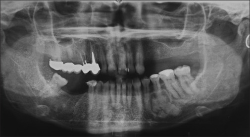
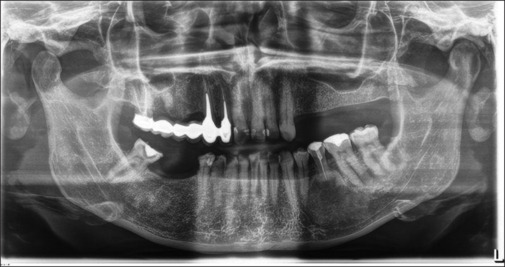
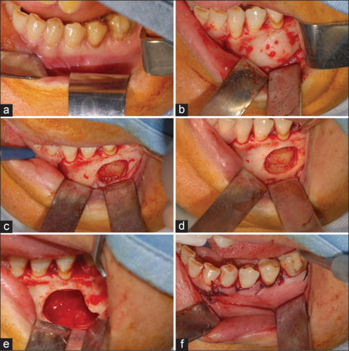
Fibrous dysplasia is a rare genetic syndrome that affects bone tissue. This pathology replaces the mineralized matrix of the bone affected with connective and fibrous tissue. This article describes a mandibular fibrous osseous dysplasia case and its surgical treatment. A 45-year-old woman complained about a slow development of swelling of the left mandibular bone. The orthopantomography (OPT) and the cone beam computed tomography (CBCT) revealed a well-circumscribed sclerotic lesion with a ground-glass appearance apical to the 3.5 element. The surgery was performed to excise the lesion. Anatomopathological examination of tissue confirmed the suspects among the diagnosis of fibrous dysplasia. The patient underwent to follow-up of 4 years, and no recurrences were found. In the absence of a univocal consensus on therapy, surgery remains the treatment of choice for unifocal forms.

 求助内容:
求助内容: 应助结果提醒方式:


