Santiago Tello-Mijares, Francisco Flores, Fomuy Woo
{"title":"Classification of Severity of Lung Parenchyma Using Saliency and Discrete Cosine Transform Energy in Computed Tomography of Patients With COVID-19.","authors":"Santiago Tello-Mijares, Francisco Flores, Fomuy Woo","doi":"10.1155/ijta/4420410","DOIUrl":null,"url":null,"abstract":"<p><p>This study proposes an automated system for assessing lung damage severity in coronavirus disease 2019 (COVID-19) patients using computed tomography (CT) images. These preprocessed CT images identify the extent of pulmonary parenchyma (PP) and ground-glass opacity and pulmonary infiltrates (GGO-PIs). Two types of images-saliency (<i>Q</i>) image and discrete cosine transform (DCT) energy image-were generated from these images. A final fused (FF) image combining <i>Q</i> and DCT of PP and GGO-PI images was then obtained. Five convolutional neural networks (CNNs) and five classic classification techniques, trained using FF and grayscale PP images, were tested. Our study is aimed at showing that a CNN model, with preprocessed images as input, has significant advantages over grayscale images. Previous work in this field primarily focused on grayscale images, which presented some limitations. This paper demonstrates how optimal results can be obtained by using the FF image rather than just the grayscale PP image. As a result, CNN models outperformed traditional artificial intelligence classification techniques. Of these, Vgg16Net performed best, delivering top-tier classification results for COVID-19 severity assessment, with a recall rate of 95.38%, precision of 96%, accuracy of 95.84%, and area under the receiver operating characteristic (AUROC) curve of 0.9585; in addition, the Vgg16Net delivers the lowest false negative (FN) results. The dataset, comprising 44 COVID-19 patients, was split equally, with half used for training and half for testing.</p>","PeriodicalId":45630,"journal":{"name":"International Journal of Telemedicine and Applications","volume":"2025 ","pages":"4420410"},"PeriodicalIF":2.2000,"publicationDate":"2025-01-06","publicationTypes":"Journal Article","fieldsOfStudy":null,"isOpenAccess":false,"openAccessPdf":"https://www.ncbi.nlm.nih.gov/pmc/articles/PMC11729514/pdf/","citationCount":"0","resultStr":null,"platform":"Semanticscholar","paperid":null,"PeriodicalName":"International Journal of Telemedicine and Applications","FirstCategoryId":"1085","ListUrlMain":"https://doi.org/10.1155/ijta/4420410","RegionNum":0,"RegionCategory":null,"ArticlePicture":[],"TitleCN":null,"AbstractTextCN":null,"PMCID":null,"EPubDate":"2025/1/1 0:00:00","PubModel":"eCollection","JCR":"Q2","JCRName":"HEALTH CARE SCIENCES & SERVICES","Score":null,"Total":0}
引用次数: 0
Abstract
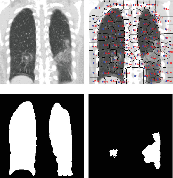
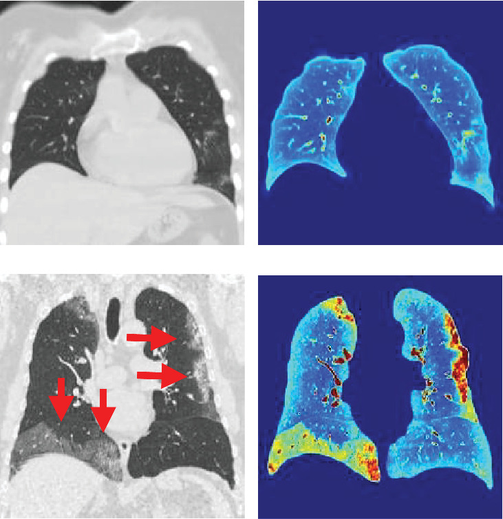
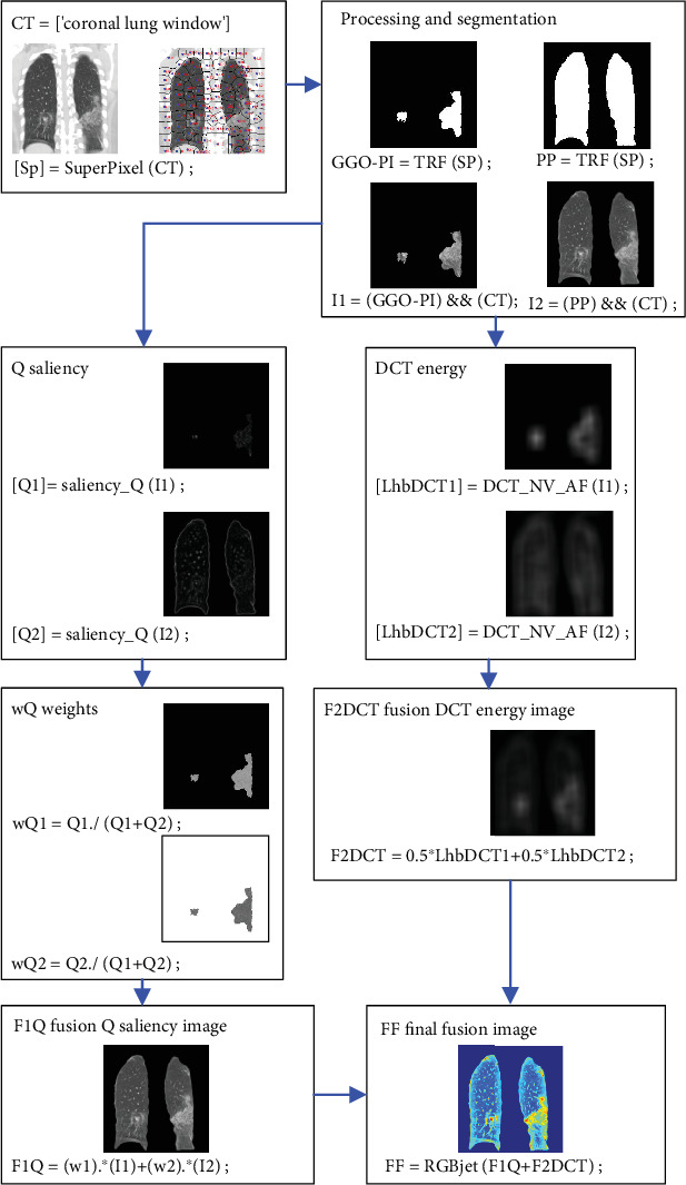
This study proposes an automated system for assessing lung damage severity in coronavirus disease 2019 (COVID-19) patients using computed tomography (CT) images. These preprocessed CT images identify the extent of pulmonary parenchyma (PP) and ground-glass opacity and pulmonary infiltrates (GGO-PIs). Two types of images-saliency (Q) image and discrete cosine transform (DCT) energy image-were generated from these images. A final fused (FF) image combining Q and DCT of PP and GGO-PI images was then obtained. Five convolutional neural networks (CNNs) and five classic classification techniques, trained using FF and grayscale PP images, were tested. Our study is aimed at showing that a CNN model, with preprocessed images as input, has significant advantages over grayscale images. Previous work in this field primarily focused on grayscale images, which presented some limitations. This paper demonstrates how optimal results can be obtained by using the FF image rather than just the grayscale PP image. As a result, CNN models outperformed traditional artificial intelligence classification techniques. Of these, Vgg16Net performed best, delivering top-tier classification results for COVID-19 severity assessment, with a recall rate of 95.38%, precision of 96%, accuracy of 95.84%, and area under the receiver operating characteristic (AUROC) curve of 0.9585; in addition, the Vgg16Net delivers the lowest false negative (FN) results. The dataset, comprising 44 COVID-19 patients, was split equally, with half used for training and half for testing.
期刊介绍:
The overall aim of the International Journal of Telemedicine and Applications is to bring together science and applications of medical practice and medical care at a distance as well as their supporting technologies such as, computing, communications, and networking technologies with emphasis on telemedicine techniques and telemedicine applications. It is directed at practicing engineers, academic researchers, as well as doctors, nurses, etc. Telemedicine is an information technology that enables doctors to perform medical consultations, diagnoses, and treatments, as well as medical education, away from patients. For example, doctors can remotely examine patients via remote viewing monitors and sound devices, and/or sampling physiological data using telecommunication. Telemedicine technology is applied to areas of emergency healthcare, videoconsulting, telecardiology, telepathology, teledermatology, teleophthalmology, teleoncology, telepsychiatry, teledentistry, etc. International Journal of Telemedicine and Applications will highlight the continued growth and new challenges in telemedicine, applications, and their supporting technologies, for both application development and basic research. Papers should emphasize original results or case studies relating to the theory and/or applications of telemedicine. Tutorial papers, especially those emphasizing multidisciplinary views of telemedicine, are also welcome. International Journal of Telemedicine and Applications employs a paperless, electronic submission and evaluation system to promote a rapid turnaround in the peer-review process.

 求助内容:
求助内容: 应助结果提醒方式:


