Teenie Kwan Tung Wong, Kenny Yat Hong Kwan, Jason Pui Yin Cheung, Kenneth Man Chee Cheung
{"title":"A case series in vertebral body tethering results in improvement in coronal Cobb angle but deterioration in axial rotation: a 3-dimensional analysis.","authors":"Teenie Kwan Tung Wong, Kenny Yat Hong Kwan, Jason Pui Yin Cheung, Kenneth Man Chee Cheung","doi":"10.21037/jss-24-59","DOIUrl":null,"url":null,"abstract":"<p><strong>Background: </strong>Vertebral body tethering (VBT) has shown improvements in coronal and sagittal plane correction in adolescent idiopathic scoliosis (AIS) patients, but axial correction over time remains unexplored. Three-dimensional (3D) spine reconstruction was used to analyse correctional changes in all spinal planes post VBT surgery.</p><p><strong>Case description: </strong>AIS subjects who underwent thoracic VBT surgery with a minimum 2-year follow-up were assessed. Biplanar radiographs were used for 3D spinal reconstructions, 3D coronal, sagittal thoracic kyphosis (TK), lumbar lordosis (LL), and axial rotation measurements were compared at pre-operative (pre-op), immediate post-operative (post-op), 1-year, and 2-year follow-up. Eight patients (7 females, 1 male) with a mean age of 11.8±1.3 years with right thoracic curves (mean 50.4°±8.1°) were followed for 26.8±4.1 months. Mean coronal Cobb angle showed significant improvement: 28.4°, 19.2°, and 27.1° at post-op, 1-year, and 2-year follow-up (P<0.001). Minimal changes were seen in sagittal plane: TK-35.2°, 39.0°, 31.3°, 37.0°; LL-46.1°, 42.8°, 36.5°, 42.8° (pre-op, post-op, 1-year, 2-year) respectively. Apical axial rotation improved from -5.5°±5.0° to -1.4°±4.8° post-op, then deteriorated to -3.2°±4.9° at 1 year and -7.0°±5.9° at 2 years, with no significant changes.</p><p><strong>Conclusions: </strong>This is the first case series to use 3D radiographic digital measurements to reveal apical axial rotation progression in thoracic curves despite improved coronal curvature. While larger scales studies with longer follow-up are needed to verify our findings, surgeons and patients should be aware of such findings in their decision to select VBT as their procedure of choice.</p>","PeriodicalId":17131,"journal":{"name":"Journal of spine surgery","volume":"10 4","pages":"687-696"},"PeriodicalIF":0.0000,"publicationDate":"2024-12-20","publicationTypes":"Journal Article","fieldsOfStudy":null,"isOpenAccess":false,"openAccessPdf":"https://www.ncbi.nlm.nih.gov/pmc/articles/PMC11732316/pdf/","citationCount":"0","resultStr":null,"platform":"Semanticscholar","paperid":null,"PeriodicalName":"Journal of spine surgery","FirstCategoryId":"1085","ListUrlMain":"https://doi.org/10.21037/jss-24-59","RegionNum":0,"RegionCategory":null,"ArticlePicture":[],"TitleCN":null,"AbstractTextCN":null,"PMCID":null,"EPubDate":"2024/11/13 0:00:00","PubModel":"Epub","JCR":"Q1","JCRName":"Medicine","Score":null,"Total":0}
引用次数: 0
Abstract
Background: Vertebral body tethering (VBT) has shown improvements in coronal and sagittal plane correction in adolescent idiopathic scoliosis (AIS) patients, but axial correction over time remains unexplored. Three-dimensional (3D) spine reconstruction was used to analyse correctional changes in all spinal planes post VBT surgery.
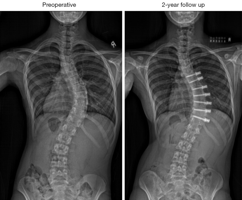
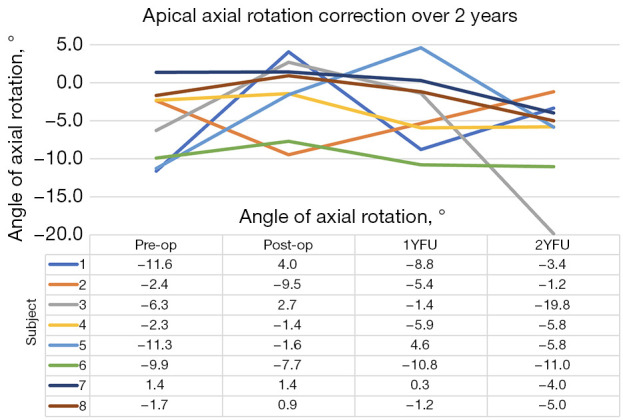
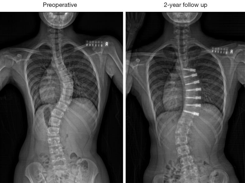
Case description: AIS subjects who underwent thoracic VBT surgery with a minimum 2-year follow-up were assessed. Biplanar radiographs were used for 3D spinal reconstructions, 3D coronal, sagittal thoracic kyphosis (TK), lumbar lordosis (LL), and axial rotation measurements were compared at pre-operative (pre-op), immediate post-operative (post-op), 1-year, and 2-year follow-up. Eight patients (7 females, 1 male) with a mean age of 11.8±1.3 years with right thoracic curves (mean 50.4°±8.1°) were followed for 26.8±4.1 months. Mean coronal Cobb angle showed significant improvement: 28.4°, 19.2°, and 27.1° at post-op, 1-year, and 2-year follow-up (P<0.001). Minimal changes were seen in sagittal plane: TK-35.2°, 39.0°, 31.3°, 37.0°; LL-46.1°, 42.8°, 36.5°, 42.8° (pre-op, post-op, 1-year, 2-year) respectively. Apical axial rotation improved from -5.5°±5.0° to -1.4°±4.8° post-op, then deteriorated to -3.2°±4.9° at 1 year and -7.0°±5.9° at 2 years, with no significant changes.
Conclusions: This is the first case series to use 3D radiographic digital measurements to reveal apical axial rotation progression in thoracic curves despite improved coronal curvature. While larger scales studies with longer follow-up are needed to verify our findings, surgeons and patients should be aware of such findings in their decision to select VBT as their procedure of choice.

 求助内容:
求助内容: 应助结果提醒方式:


