Seung Yong Shin, Min Soo Cho, Jinhoon Nam, Jie-Hyun Kim, Young Hoon Yoon, Hyojin Park, Jeonghyun Kang, Jae Jun Park
{"title":"Clinical outcomes and risk factors of post-polypectomy microperforation in patients with colorectal neoplasia: a case-control study.","authors":"Seung Yong Shin, Min Soo Cho, Jinhoon Nam, Jie-Hyun Kim, Young Hoon Yoon, Hyojin Park, Jeonghyun Kang, Jae Jun Park","doi":"10.1177/26317745241312521","DOIUrl":null,"url":null,"abstract":"<p><strong>Background: </strong>Colonoscopic polypectomy significantly reduces the incidence of colorectal cancer, but it carries potential risks, with colonic perforation being the most common and associated with significant morbidity.</p><p><strong>Objectives: </strong>This study evaluated the clinical outcomes and risk factors of microperforation during colonoscopic polypectomy.</p><p><strong>Design: </strong>A retrospective cohort study.</p><p><strong>Methods: </strong>We retrospectively reviewed the patients' records who underwent colonoscopic polypectomy and subsequent plain radiographic examination to monitor perforation. Patients with pneumoperitoneum detected on plain radiography were enrolled. Patients who underwent adverse event-free colonoscopic polypectomies within 1 week of each case and were matched 2:1 by age and sex to the cases were selected as controls.</p><p><strong>Results: </strong>Microperforations occurred in 12 patients (8 males; age: median 64.5 years). Polyps with microperforations were more frequent in the right colon (83.3% vs 33.3%). Endoscopic mucosal resection with precutting (EMR-P; 16.7% vs 0.0%) or hot-snare polypectomy (8.3% vs 0.0%) was more frequently performed in the microperforation group. Muscle fibers at the polypectomy site were more frequently visible in the microperforation group (58.3% vs 8.3%). By multivariate analysis, right colon location and visible muscle fibers were independent risk factors for microperforation. All patients with microperforation received intravenous antibiotics and were advised to fast. Patients responded well to these conservative treatments and were discharged after a median of 3 (2-6.75) days of hospital stay.</p><p><strong>Conclusion: </strong>Our data suggest that conservative treatment is feasible and could be the primary management option for selected patients with microperforations postcolonoscopic polypectomy. Right-sided colonic polyps and visible muscle fibers predispose to microperforations.</p>","PeriodicalId":40947,"journal":{"name":"Therapeutic Advances in Gastrointestinal Endoscopy","volume":"18 ","pages":"26317745241312521"},"PeriodicalIF":2.4000,"publicationDate":"2025-01-09","publicationTypes":"Journal Article","fieldsOfStudy":null,"isOpenAccess":false,"openAccessPdf":"https://www.ncbi.nlm.nih.gov/pmc/articles/PMC11719433/pdf/","citationCount":"0","resultStr":null,"platform":"Semanticscholar","paperid":null,"PeriodicalName":"Therapeutic Advances in Gastrointestinal Endoscopy","FirstCategoryId":"1085","ListUrlMain":"https://doi.org/10.1177/26317745241312521","RegionNum":0,"RegionCategory":null,"ArticlePicture":[],"TitleCN":null,"AbstractTextCN":null,"PMCID":null,"EPubDate":"2025/1/1 0:00:00","PubModel":"eCollection","JCR":"Q2","JCRName":"GASTROENTEROLOGY & HEPATOLOGY","Score":null,"Total":0}
引用次数: 0
Abstract
Background: Colonoscopic polypectomy significantly reduces the incidence of colorectal cancer, but it carries potential risks, with colonic perforation being the most common and associated with significant morbidity.
Objectives: This study evaluated the clinical outcomes and risk factors of microperforation during colonoscopic polypectomy.
Design: A retrospective cohort study.
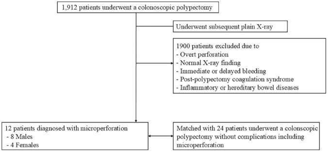
Methods: We retrospectively reviewed the patients' records who underwent colonoscopic polypectomy and subsequent plain radiographic examination to monitor perforation. Patients with pneumoperitoneum detected on plain radiography were enrolled. Patients who underwent adverse event-free colonoscopic polypectomies within 1 week of each case and were matched 2:1 by age and sex to the cases were selected as controls.
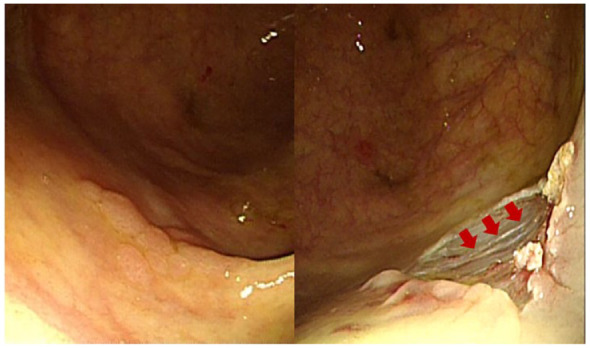
Results: Microperforations occurred in 12 patients (8 males; age: median 64.5 years). Polyps with microperforations were more frequent in the right colon (83.3% vs 33.3%). Endoscopic mucosal resection with precutting (EMR-P; 16.7% vs 0.0%) or hot-snare polypectomy (8.3% vs 0.0%) was more frequently performed in the microperforation group. Muscle fibers at the polypectomy site were more frequently visible in the microperforation group (58.3% vs 8.3%). By multivariate analysis, right colon location and visible muscle fibers were independent risk factors for microperforation. All patients with microperforation received intravenous antibiotics and were advised to fast. Patients responded well to these conservative treatments and were discharged after a median of 3 (2-6.75) days of hospital stay.
Conclusion: Our data suggest that conservative treatment is feasible and could be the primary management option for selected patients with microperforations postcolonoscopic polypectomy. Right-sided colonic polyps and visible muscle fibers predispose to microperforations.
背景:结肠镜下息肉切除术可显著降低结直肠癌的发病率,但也存在潜在风险,结肠穿孔最为常见,且发病率较高。目的:评价结肠镜下息肉切除术中微穿孔的临床结局及危险因素。设计:回顾性队列研究。方法:我们回顾性地回顾了接受结肠镜息肉切除术和随后的x线平片检查以监测穿孔的患者记录。在x线平片上发现气腹的患者被纳入研究。选取在每个病例1周内接受无不良事件结肠镜息肉切除术的患者作为对照,并按年龄和性别2:1匹配。结果:12例患者出现微穿孔,其中男性8例;年龄:中位64.5岁)。右结肠息肉伴微穿孔发生率更高(83.3% vs 33.3%)。内镜下粘膜预切切除术(EMR-P);(16.7% vs 0.0%)或热圈套息肉切除术(8.3% vs 0.0%)在微穿孔组更常见。微穿孔组息肉切除部位的肌纤维更常见(58.3% vs 8.3%)。多因素分析显示,右结肠位置和可见肌纤维是微穿孔的独立危险因素。所有微穿孔患者均静脉注射抗生素,并建议禁食。患者对这些保守治疗反应良好,中位住院3(2-6.75)天后出院。结论:我们的数据表明保守治疗是可行的,可以作为结肠镜息肉切除术后微穿孔患者的主要治疗选择。右侧结肠息肉和可见肌纤维易导致微穿孔。

 求助内容:
求助内容: 应助结果提醒方式:


