{"title":"Resection of shrinking secondary thymic cyst during follow-up-a case report.","authors":"Taekyung Kang, Mi-Jin Kang","doi":"10.21037/med-24-13","DOIUrl":null,"url":null,"abstract":"<p><strong>Background: </strong>Thymic cysts can be classified as congenital or acquired. Most thymic cysts do not change in size over a short period of time. Although very rare, thymic cyst rupture is associated with serious complications, such as mediastinal hemorrhage and hemothorax. We experienced a case of partial rupture of a secondary thymic cyst, in an asymptomatic patient.</p><p><strong>Case description: </strong>A 60-year-old woman visited Inje University Sanggye Paik Hospital with left hilar bulging detected on routine chest radiograph. A chest computed tomography (CT) scan revealed a 6 cm well-defined cystic mass with partial septation in the prevascular mediastinum. Thus, secondary thymic cyst was suggested. On the follow-up chest CT scan taken 3 months later, the size of the thymic cyst decreased, while the solid portion increased slightly, suggesting the potential presence of malignancy. Consequently, surgery was conducted. Adhesion to the lung and aorta was observed, but they were relatively well separated. The pathological findings revealed a partially ruptured thymic cyst with fat necrosis and multifocal granulomas.</p><p><strong>Conclusions: </strong>There are controversies in the treatment of thymic cysts. Some clinicians prefer strict medical supervision to avoid unnecessary surgery, while others advocate immediate excision to avoid complication. However, if any changes are observed during the follow-up of the thymic cyst, it may indicate malignant transformation or rupture, necessitating prompt surgical excision.</p>","PeriodicalId":74139,"journal":{"name":"Mediastinum (Hong Kong, China)","volume":"8 ","pages":"52"},"PeriodicalIF":0.0000,"publicationDate":"2024-07-12","publicationTypes":"Journal Article","fieldsOfStudy":null,"isOpenAccess":false,"openAccessPdf":"https://www.ncbi.nlm.nih.gov/pmc/articles/PMC11707434/pdf/","citationCount":"0","resultStr":null,"platform":"Semanticscholar","paperid":null,"PeriodicalName":"Mediastinum (Hong Kong, China)","FirstCategoryId":"1085","ListUrlMain":"https://doi.org/10.21037/med-24-13","RegionNum":0,"RegionCategory":null,"ArticlePicture":[],"TitleCN":null,"AbstractTextCN":null,"PMCID":null,"EPubDate":"2024/1/1 0:00:00","PubModel":"eCollection","JCR":"","JCRName":"","Score":null,"Total":0}
引用次数: 0
Abstract
Background: Thymic cysts can be classified as congenital or acquired. Most thymic cysts do not change in size over a short period of time. Although very rare, thymic cyst rupture is associated with serious complications, such as mediastinal hemorrhage and hemothorax. We experienced a case of partial rupture of a secondary thymic cyst, in an asymptomatic patient.
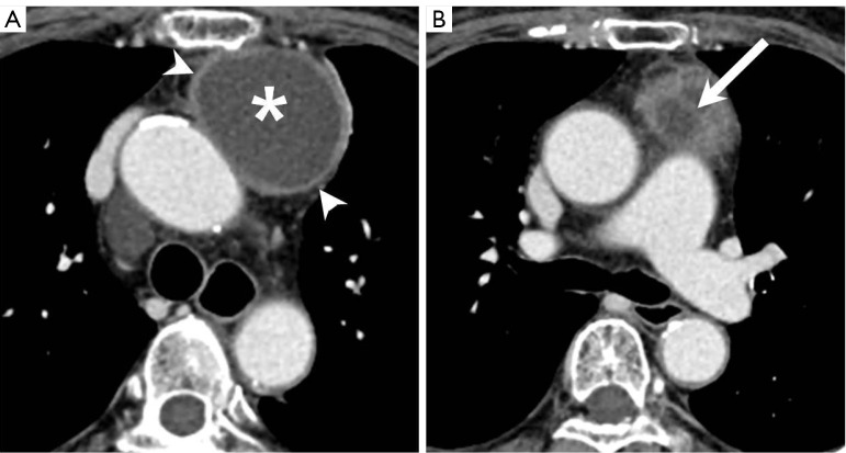
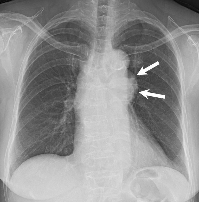
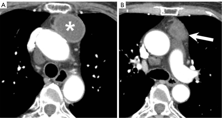
Case description: A 60-year-old woman visited Inje University Sanggye Paik Hospital with left hilar bulging detected on routine chest radiograph. A chest computed tomography (CT) scan revealed a 6 cm well-defined cystic mass with partial septation in the prevascular mediastinum. Thus, secondary thymic cyst was suggested. On the follow-up chest CT scan taken 3 months later, the size of the thymic cyst decreased, while the solid portion increased slightly, suggesting the potential presence of malignancy. Consequently, surgery was conducted. Adhesion to the lung and aorta was observed, but they were relatively well separated. The pathological findings revealed a partially ruptured thymic cyst with fat necrosis and multifocal granulomas.
Conclusions: There are controversies in the treatment of thymic cysts. Some clinicians prefer strict medical supervision to avoid unnecessary surgery, while others advocate immediate excision to avoid complication. However, if any changes are observed during the follow-up of the thymic cyst, it may indicate malignant transformation or rupture, necessitating prompt surgical excision.

 求助内容:
求助内容: 应助结果提醒方式:


