Longitudinal Changes in Medial Meniscal Extrusion After ACL Injury and Reconstruction and Its Relationship With Cartilage Degeneration Assessed Using MRI-Based T1ρ and T2 Analysis.
Shotaro Watanabe, Gabby B Joseph, Dai Sato, Drew A Lansdown, Julio Brandao Guimaraes, Thomas M Link, Chunbong Benjamin Ma
{"title":"Longitudinal Changes in Medial Meniscal Extrusion After ACL Injury and Reconstruction and Its Relationship With Cartilage Degeneration Assessed Using MRI-Based T1ρ and T2 Analysis.","authors":"Shotaro Watanabe, Gabby B Joseph, Dai Sato, Drew A Lansdown, Julio Brandao Guimaraes, Thomas M Link, Chunbong Benjamin Ma","doi":"10.1177/03635465241305734","DOIUrl":null,"url":null,"abstract":"<p><strong>Background: </strong>Anterior cruciate ligament (ACL) injury often leads to posttraumatic osteoarthritis (PTOA), despite ACL reconstruction (ACLR). Medial meniscal extrusion (MME) is implicated in PTOA progression but remains understudied after ACL injury and ACLR.</p><p><strong>Hypothesis/purpose: </strong>It was hypothesized that MME would increase longitudinally after ACL injury and ACLR, with greater changes in the ipsilateral knee compared with the contralateral knee, leading to cartilage degeneration. The study aimed to assess MME 3 years after ACLR and its relationship with magnetic resonance imaging (MRI) T1ρ and T2 as cartilage degeneration markers.</p><p><strong>Study design: </strong>Cohort study; Level of evidence, 2.</p><p><strong>Methods: </strong>MME and relative percentage of extrusion (RPE) were measured on 3 coronal slices of 3-dimensional fast spin-echo images and the mean values were used. T1ρ and T2 sequences were obtained and cartilage compositional measurements were performed using in-house developed software with MATLAB. Mixed models were used to assess the longitudinal changes and linear regression was used to assess the relationships between RPE and T1ρ and T2 values.</p><p><strong>Results: </strong>A total of 54 participants with unilateral ACL injuries underwent preoperative bilateral knee MRI. A total of 36 participants completed MR scans at 6 months and 3 years after ACLR. MME and RPE measurements demonstrated high reliability (ICC > 0.88 and > 0.91, respectively). The predicted values of MME and RPE from the mixed models showed that the ipsilateral side had significantly greater MME and RPE than the contralateral side at all 3 time points (<i>P</i> = .023 for MME; <i>P</i> = .013 for RPE at baseline; and <i>P</i> < .001 at 6 months and <i>P</i> < .001 at 3 years for both MME and RPE). The rate of change of MME and RPE on the ipsilateral side was significantly greater than that on the contralateral side (<i>P</i> < .001). Postoperative RPE was associated with T1ρ and T2 values in the posterior medial femoral condyle.</p><p><strong>Conclusion: </strong>MME and RPE obtained pre- and postoperatively after ACLR on the ipsilateral side were significantly greater than those on the contralateral side, and the longitudinal increases on the ipsilateral side were greater than those on the contralateral side. Postoperative RPE was significantly associated with cartilage degeneration in the posterior medial femoral condyle.</p>","PeriodicalId":55528,"journal":{"name":"American Journal of Sports Medicine","volume":" ","pages":"350-359"},"PeriodicalIF":4.5000,"publicationDate":"2025-02-01","publicationTypes":"Journal Article","fieldsOfStudy":null,"isOpenAccess":false,"openAccessPdf":"https://www.ncbi.nlm.nih.gov/pmc/articles/PMC11796289/pdf/","citationCount":"0","resultStr":null,"platform":"Semanticscholar","paperid":null,"PeriodicalName":"American Journal of Sports Medicine","FirstCategoryId":"3","ListUrlMain":"https://doi.org/10.1177/03635465241305734","RegionNum":1,"RegionCategory":"医学","ArticlePicture":[],"TitleCN":null,"AbstractTextCN":null,"PMCID":null,"EPubDate":"2025/1/2 0:00:00","PubModel":"Epub","JCR":"Q1","JCRName":"ORTHOPEDICS","Score":null,"Total":0}
引用次数: 0
Abstract
Background: Anterior cruciate ligament (ACL) injury often leads to posttraumatic osteoarthritis (PTOA), despite ACL reconstruction (ACLR). Medial meniscal extrusion (MME) is implicated in PTOA progression but remains understudied after ACL injury and ACLR.
Hypothesis/purpose: It was hypothesized that MME would increase longitudinally after ACL injury and ACLR, with greater changes in the ipsilateral knee compared with the contralateral knee, leading to cartilage degeneration. The study aimed to assess MME 3 years after ACLR and its relationship with magnetic resonance imaging (MRI) T1ρ and T2 as cartilage degeneration markers.
Study design: Cohort study; Level of evidence, 2.
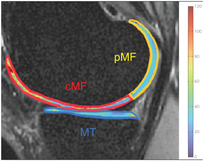
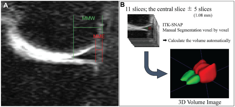
Methods: MME and relative percentage of extrusion (RPE) were measured on 3 coronal slices of 3-dimensional fast spin-echo images and the mean values were used. T1ρ and T2 sequences were obtained and cartilage compositional measurements were performed using in-house developed software with MATLAB. Mixed models were used to assess the longitudinal changes and linear regression was used to assess the relationships between RPE and T1ρ and T2 values.
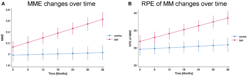
Results: A total of 54 participants with unilateral ACL injuries underwent preoperative bilateral knee MRI. A total of 36 participants completed MR scans at 6 months and 3 years after ACLR. MME and RPE measurements demonstrated high reliability (ICC > 0.88 and > 0.91, respectively). The predicted values of MME and RPE from the mixed models showed that the ipsilateral side had significantly greater MME and RPE than the contralateral side at all 3 time points (P = .023 for MME; P = .013 for RPE at baseline; and P < .001 at 6 months and P < .001 at 3 years for both MME and RPE). The rate of change of MME and RPE on the ipsilateral side was significantly greater than that on the contralateral side (P < .001). Postoperative RPE was associated with T1ρ and T2 values in the posterior medial femoral condyle.
Conclusion: MME and RPE obtained pre- and postoperatively after ACLR on the ipsilateral side were significantly greater than those on the contralateral side, and the longitudinal increases on the ipsilateral side were greater than those on the contralateral side. Postoperative RPE was significantly associated with cartilage degeneration in the posterior medial femoral condyle.
期刊介绍:
An invaluable resource for the orthopaedic sports medicine community, _The American Journal of Sports Medicine_ is a peer-reviewed scientific journal, first published in 1972. It is the official publication of the [American Orthopaedic Society for Sports Medicine (AOSSM)](http://www.sportsmed.org/)! The journal acts as an important forum for independent orthopaedic sports medicine research and education, allowing clinical practitioners the ability to make decisions based on sound scientific information.
This journal is a must-read for:
* Orthopaedic Surgeons and Specialists
* Sports Medicine Physicians
* Physiatrists
* Athletic Trainers
* Team Physicians
* And Physical Therapists
 求助内容:
求助内容: 应助结果提醒方式:


