{"title":"The Role of In Vivo Confocal Microscopy in Ocular Allergies.","authors":"Cem Şimşek, Takashi Kojima, Murat Doğru","doi":"10.4274/tjo.galenos.2024.49769","DOIUrl":null,"url":null,"abstract":"<p><p>In vivo confocal microscopy (IVCM) is a non-invasive imaging technique used to visualize the layers of the cornea and conjunctiva in real time. In patients with atopic keratoconjunctivitis (AKC) and vernal keratoconjunctivitis (VKC), this technology can be useful in diagnosing and monitoring the disease, as well as evaluating the efficacy of treatments. IVCM can reveal subclinical abnormalities in the corneal and conjunctival epithelium such as inflammatory cell infiltrates and tissue damage, which can provide insight into the pathogenesis of AKC. In AKC, IVCM reveals changes around the conjunctival papillae, inflammatory cells around punctate defects in the corneal epithelium, changes in subbasal nerve morphology, and deteriorations in the goblet cells in the meibomian gland are observed. In VKC, alterations can be observed in the diameter, brightness and nucleus/cytoplasm ratio of the superficial epithelial cells in the cornea. The use of IVCM in AKC and VKC can therefore aid in the early detection and management of the disease, as well as contribute to a better understanding of its underlying mechanisms.</p>","PeriodicalId":23373,"journal":{"name":"Turkish Journal of Ophthalmology","volume":"54 6","pages":"344-353"},"PeriodicalIF":0.0000,"publicationDate":"2024-12-31","publicationTypes":"Journal Article","fieldsOfStudy":null,"isOpenAccess":false,"openAccessPdf":"https://www.ncbi.nlm.nih.gov/pmc/articles/PMC11707458/pdf/","citationCount":"0","resultStr":null,"platform":"Semanticscholar","paperid":null,"PeriodicalName":"Turkish Journal of Ophthalmology","FirstCategoryId":"1085","ListUrlMain":"https://doi.org/10.4274/tjo.galenos.2024.49769","RegionNum":0,"RegionCategory":null,"ArticlePicture":[],"TitleCN":null,"AbstractTextCN":null,"PMCID":null,"EPubDate":"","PubModel":"","JCR":"Q3","JCRName":"Medicine","Score":null,"Total":0}
引用次数: 0
Abstract
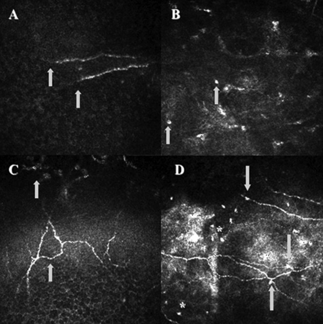
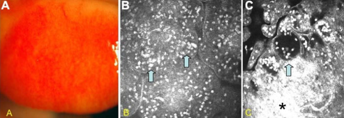
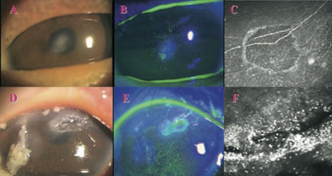
In vivo confocal microscopy (IVCM) is a non-invasive imaging technique used to visualize the layers of the cornea and conjunctiva in real time. In patients with atopic keratoconjunctivitis (AKC) and vernal keratoconjunctivitis (VKC), this technology can be useful in diagnosing and monitoring the disease, as well as evaluating the efficacy of treatments. IVCM can reveal subclinical abnormalities in the corneal and conjunctival epithelium such as inflammatory cell infiltrates and tissue damage, which can provide insight into the pathogenesis of AKC. In AKC, IVCM reveals changes around the conjunctival papillae, inflammatory cells around punctate defects in the corneal epithelium, changes in subbasal nerve morphology, and deteriorations in the goblet cells in the meibomian gland are observed. In VKC, alterations can be observed in the diameter, brightness and nucleus/cytoplasm ratio of the superficial epithelial cells in the cornea. The use of IVCM in AKC and VKC can therefore aid in the early detection and management of the disease, as well as contribute to a better understanding of its underlying mechanisms.
期刊介绍:
The Turkish Journal of Ophthalmology (TJO) is the only scientific periodical publication of the Turkish Ophthalmological Association and has been published since January 1929. In its early years, the journal was published in Turkish and French. Although there were temporary interruptions in the publication of the journal due to various challenges, the Turkish Journal of Ophthalmology has been published continually from 1971 to the present. The target audience includes specialists and physicians in training in ophthalmology in all relevant disciplines.

 求助内容:
求助内容: 应助结果提醒方式:


