A Rare and Intriguing Case of Papillary Thyroid Carcinoma with Tumor Thrombus Extending into the Right Ventricle: Documentation with 131 I-NaI-SPECT/CT, MRI, and 18 F-FDG-PET/CT.
IF 0.9 Q4 RADIOLOGY, NUCLEAR MEDICINE & MEDICAL IMAGING
{"title":"A Rare and Intriguing Case of Papillary Thyroid Carcinoma with Tumor Thrombus Extending into the Right Ventricle: Documentation with <sup>131</sup> I-NaI-SPECT/CT, MRI, and <sup>18</sup> F-FDG-PET/CT.","authors":"Parth Baberwal, Ramesh Asopa, Sandip Basu","doi":"10.1055/s-0044-1788737","DOIUrl":null,"url":null,"abstract":"<p><p>A unique case of papillary carcinoma of the thyroid with an extensive tumor thrombus extending into the right ventricle is presented. The patient was a known case of solid variant of papillary carcinoma of thyroid, post three cycles of radioiodine therapy, had reported for a diagnostic <sup>131</sup> I-NaI scintigraphy as a part of the workup for planning the next <sup>131</sup> I therapy. Clinically, the patient was asymptomatic. <sup>131</sup> I-NaI scintigraphy showed an arcuate pattern concentration of tracer in the upper mediastinum, which descended up to the lower mediastinum. A <sup>131</sup> I-NaI single photon emission computed tomography/computed tomography (SPECT/CT) showed a tracer avid tumor with an extensive tumor thrombus extending from the left brachiocephalic vein to the right ventricle. <sup>18</sup> F-fluorodeoxyglucose positron emission tomography/computed tomography ( <sup>18</sup> F-FDG-PET/CT) and magnetic resonance imaging (MRI) demonstrated similar findings. The patient was decided to be managed with tyrosine kinase inhibitors as surgical intervention was not deemed possible due to the involvement of major vessels and the high risk of bleeding.</p>","PeriodicalId":23742,"journal":{"name":"World Journal of Nuclear Medicine","volume":"23 4","pages":"295-298"},"PeriodicalIF":0.9000,"publicationDate":"2024-08-05","publicationTypes":"Journal Article","fieldsOfStudy":null,"isOpenAccess":false,"openAccessPdf":"https://www.ncbi.nlm.nih.gov/pmc/articles/PMC11637636/pdf/","citationCount":"0","resultStr":null,"platform":"Semanticscholar","paperid":null,"PeriodicalName":"World Journal of Nuclear Medicine","FirstCategoryId":"1085","ListUrlMain":"https://doi.org/10.1055/s-0044-1788737","RegionNum":0,"RegionCategory":null,"ArticlePicture":[],"TitleCN":null,"AbstractTextCN":null,"PMCID":null,"EPubDate":"2024/12/1 0:00:00","PubModel":"eCollection","JCR":"Q4","JCRName":"RADIOLOGY, NUCLEAR MEDICINE & MEDICAL IMAGING","Score":null,"Total":0}
引用次数: 0
Abstract
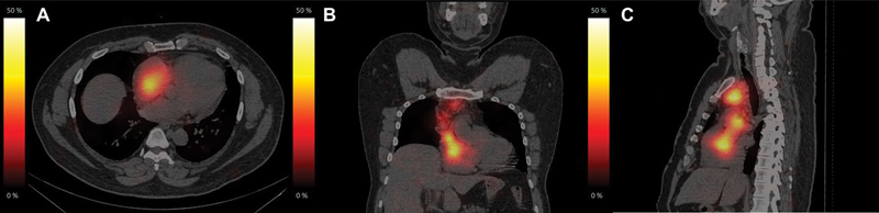
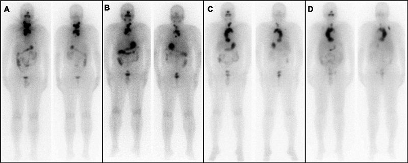
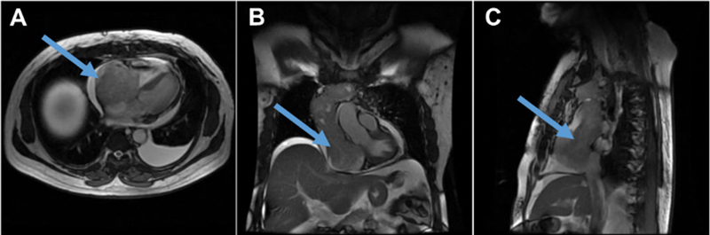
A unique case of papillary carcinoma of the thyroid with an extensive tumor thrombus extending into the right ventricle is presented. The patient was a known case of solid variant of papillary carcinoma of thyroid, post three cycles of radioiodine therapy, had reported for a diagnostic 131 I-NaI scintigraphy as a part of the workup for planning the next 131 I therapy. Clinically, the patient was asymptomatic. 131 I-NaI scintigraphy showed an arcuate pattern concentration of tracer in the upper mediastinum, which descended up to the lower mediastinum. A 131 I-NaI single photon emission computed tomography/computed tomography (SPECT/CT) showed a tracer avid tumor with an extensive tumor thrombus extending from the left brachiocephalic vein to the right ventricle. 18 F-fluorodeoxyglucose positron emission tomography/computed tomography ( 18 F-FDG-PET/CT) and magnetic resonance imaging (MRI) demonstrated similar findings. The patient was decided to be managed with tyrosine kinase inhibitors as surgical intervention was not deemed possible due to the involvement of major vessels and the high risk of bleeding.

 求助内容:
求助内容: 应助结果提醒方式:


