Diagnosis of fibrotic interstitial lung diseases based on the combination of label-free quantitative multiphoton fiber histology and machine learning.

IF 5.1 2区 医学 Q1 MEDICINE, RESEARCH & EXPERIMENTAL
Wenzhuo Qiu, Qingyang Wang, Ying Zhang, Xiuxue Cao, Ling Zhao, Longhao Cao, Yuxuan Sun, Feili Yang, Yuanyuan Guo, Yuming Sui, Ziyi Chang, Congcong Wang, Lifang Cui, Yun Niu, Pingping Liu, Jie Lin, Shixuan Liu, Jia Guo, Bei Wang, Ruiqi Zhong, Ce Wang, Wei Liu, Dawei Li, Huaping Dai, Sheng Xie, Heping Cheng, Aimin Wang, Dingrong Zhong
{"title":"Diagnosis of fibrotic interstitial lung diseases based on the combination of label-free quantitative multiphoton fiber histology and machine learning.","authors":"Wenzhuo Qiu, Qingyang Wang, Ying Zhang, Xiuxue Cao, Ling Zhao, Longhao Cao, Yuxuan Sun, Feili Yang, Yuanyuan Guo, Yuming Sui, Ziyi Chang, Congcong Wang, Lifang Cui, Yun Niu, Pingping Liu, Jie Lin, Shixuan Liu, Jia Guo, Bei Wang, Ruiqi Zhong, Ce Wang, Wei Liu, Dawei Li, Huaping Dai, Sheng Xie, Heping Cheng, Aimin Wang, Dingrong Zhong","doi":"10.1016/j.labinv.2024.102210","DOIUrl":null,"url":null,"abstract":"<p><p>Interstitial lung disease (ILD), characterized by inflammation and fibrosis, often suffers from low diagnostic accuracy and consistency. Traditional H&E staining primarily reveals cellular inflammation with limited detail on fibrosis. To address these issues, we introduce a pioneering label-free quantitative multiphoton fiber histology (MPFH) technique that delineates the intricate characteristics of collagen and elastin fibers for ILDs diagnosis. We acquired co-located multiphoton and H&E-stained images from a single tissue slice. Multiphoton imaging was performed on the deparaffinized section to obtain fibrotic tissue information, followed by H&E staining to capture cellular information. This approach was tested in a blinded diagnostic trial among 7 pathologists involving 14 relatively normal lung patients and 31 ILD patients (11 idiopathic pulmonary fibrosis (IPF) / usual interstitial pneumonia (UIP), 14 nonspecific interstitial pneumonia (NSIP), and 6 pleuroparenchymal fibroelastosis (PPFE)). A customized algorithm extracted quantitative fiber indicators from multiphoton images. These indicators, combined with clinical and radiological features, were used to develop an automatic multi-class ILDs classifier. Using MPFH, we can acquire high-quality, co-localized images of collagen fibers, elastin fibers, and cells. We found that the type, distribution, and degree of fibrotic proliferation can effectively distinguish between different subtypes. The blind study showed MPFH enhanced diagnostic consistency (kappa values from 0.56 to 0.72) and accuracy (from 73.0% to 82.5%, p=0.0090). The combination of quantitative fiber indicators effectively distinguished between different tissues, with areas under the receiver operating characteristic curves exceeding 0.92. The automatic classifier achieved 93.8% accuracy, closely paralleling the 92.2% accuracy of expert pathologists. The outcomes of our research underscore the transformative potential of MPFH in the field of f-ILD diagnostics. By integrating quantitative analysis of fiber characteristics with advanced machine learning algorithms, MPFH facilitates the automatic and accurate identification of various fibrotic disease subtypes, showcasing a significant leap forward in precision diagnostics.</p>","PeriodicalId":17930,"journal":{"name":"Laboratory Investigation","volume":" ","pages":"102210"},"PeriodicalIF":5.1000,"publicationDate":"2024-12-13","publicationTypes":"Journal Article","fieldsOfStudy":null,"isOpenAccess":false,"openAccessPdf":"","citationCount":"0","resultStr":null,"platform":"Semanticscholar","paperid":null,"PeriodicalName":"Laboratory Investigation","FirstCategoryId":"3","ListUrlMain":"https://doi.org/10.1016/j.labinv.2024.102210","RegionNum":2,"RegionCategory":"医学","ArticlePicture":[],"TitleCN":null,"AbstractTextCN":null,"PMCID":null,"EPubDate":"","PubModel":"","JCR":"Q1","JCRName":"MEDICINE, RESEARCH & EXPERIMENTAL","Score":null,"Total":0}
引用次数: 0

Abstract

Interstitial lung disease (ILD), characterized by inflammation and fibrosis, often suffers from low diagnostic accuracy and consistency. Traditional H&E staining primarily reveals cellular inflammation with limited detail on fibrosis. To address these issues, we introduce a pioneering label-free quantitative multiphoton fiber histology (MPFH) technique that delineates the intricate characteristics of collagen and elastin fibers for ILDs diagnosis. We acquired co-located multiphoton and H&E-stained images from a single tissue slice. Multiphoton imaging was performed on the deparaffinized section to obtain fibrotic tissue information, followed by H&E staining to capture cellular information. This approach was tested in a blinded diagnostic trial among 7 pathologists involving 14 relatively normal lung patients and 31 ILD patients (11 idiopathic pulmonary fibrosis (IPF) / usual interstitial pneumonia (UIP), 14 nonspecific interstitial pneumonia (NSIP), and 6 pleuroparenchymal fibroelastosis (PPFE)). A customized algorithm extracted quantitative fiber indicators from multiphoton images. These indicators, combined with clinical and radiological features, were used to develop an automatic multi-class ILDs classifier. Using MPFH, we can acquire high-quality, co-localized images of collagen fibers, elastin fibers, and cells. We found that the type, distribution, and degree of fibrotic proliferation can effectively distinguish between different subtypes. The blind study showed MPFH enhanced diagnostic consistency (kappa values from 0.56 to 0.72) and accuracy (from 73.0% to 82.5%, p=0.0090). The combination of quantitative fiber indicators effectively distinguished between different tissues, with areas under the receiver operating characteristic curves exceeding 0.92. The automatic classifier achieved 93.8% accuracy, closely paralleling the 92.2% accuracy of expert pathologists. The outcomes of our research underscore the transformative potential of MPFH in the field of f-ILD diagnostics. By integrating quantitative analysis of fiber characteristics with advanced machine learning algorithms, MPFH facilitates the automatic and accurate identification of various fibrotic disease subtypes, showcasing a significant leap forward in precision diagnostics.

基于无标记定量多光子纤维组织学和机器学习相结合的纤维化间质性肺疾病诊断。
以炎症和纤维化为特征的间质性肺病(ILD)通常诊断准确性和一致性较低。传统的 H&E 染色主要显示的是细胞炎症,对纤维化的细节了解有限。为了解决这些问题,我们引入了一种开创性的无标记定量多光子纤维组织学(MPFH)技术,该技术能描绘出胶原纤维和弹性纤维的复杂特征,用于诊断 ILDs。我们从单个组织切片中获取共定位的多光子和H&E染色图像。对去石蜡切片进行多光子成像以获取纤维组织信息,然后进行H&E染色以获取细胞信息。7 位病理学家对这种方法进行了盲法诊断试验,其中包括 14 位相对正常的肺部患者和 31 位 ILD 患者(11 位特发性肺纤维化 (IPF) / 常发性间质性肺炎 (UIP)、14 位非特异性间质性肺炎 (NSIP) 和 6 位胸膜间质纤维细胞增生症 (PPFE))。一种定制算法可从多光子图像中提取定量纤维指标。这些指标与临床和放射学特征相结合,用于开发多类 ILDs 自动分类器。利用多光子成像技术,我们可以获得高质量的胶原纤维、弹性纤维和细胞的共定位图像。我们发现,纤维增生的类型、分布和程度可以有效区分不同的亚型。盲法研究显示,MPFH 提高了诊断一致性(kappa 值从 0.56 到 0.72)和准确性(从 73.0% 到 82.5%,p=0.0090)。纤维定量指标的组合可有效区分不同组织,接收者操作特征曲线下的面积超过 0.92。自动分类器的准确率达到 93.8%,与病理专家 92.2% 的准确率相当。我们的研究成果凸显了 MPFH 在 f-ILD 诊断领域的变革潜力。通过将纤维特征的定量分析与先进的机器学习算法相结合,MPFH有助于自动、准确地识别各种纤维化疾病亚型,展示了精准诊断领域的重大飞跃。
本文章由计算机程序翻译,如有差异,请以英文原文为准。
求助全文
约1分钟内获得全文 求助全文
来源期刊
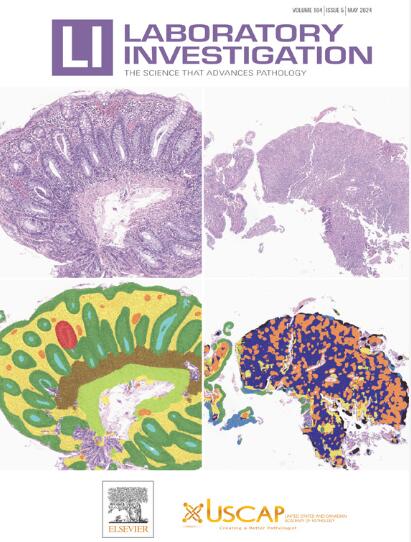
Laboratory Investigation
Laboratory Investigation 医学-病理学
CiteScore
8.30
自引率
0.00%
发文量
125
审稿时长
2 months
期刊介绍: Laboratory Investigation is an international journal owned by the United States and Canadian Academy of Pathology. Laboratory Investigation offers prompt publication of high-quality original research in all biomedical disciplines relating to the understanding of human disease and the application of new methods to the diagnosis of disease. Both human and experimental studies are welcome.
×
引用
GB/T 7714-2015
复制
MLA
复制
APA
复制
导出至
BibTeX EndNote RefMan NoteFirst NoteExpress
×
提示
您的信息不完整,为了账户安全,请先补充。
现在去补充
×
提示
您因"违规操作"
具体请查看互助需知
我知道了
×
提示
确定
请完成安全验证×
copy
已复制链接
快去分享给好友吧!
我知道了
右上角分享
点击右上角分享
0
联系我们:info@booksci.cn Book学术提供免费学术资源搜索服务,方便国内外学者检索中英文文献。致力于提供最便捷和优质的服务体验。 Copyright © 2023 布克学术 All rights reserved.
京ICP备2023020795号-1
ghs 京公网安备 11010802042870号
Book学术文献互助
Book学术文献互助群
群 号:481959085
Book学术官方微信