Stephen Rankin, Caitlin Fountain, Alastair J Gemmell, Daire Quinn, Alasdair Henderson, John McClure, Sandy Small, Balaji Venugopal, Pamela McKay, Piotr J Slomka, David Colville, Mark C Petrie, Giselle C Meléndez, Ninian N Lang
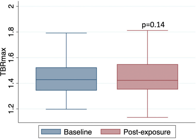
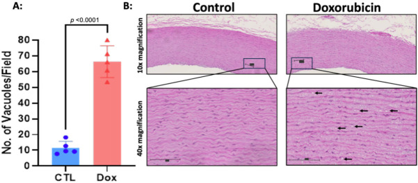
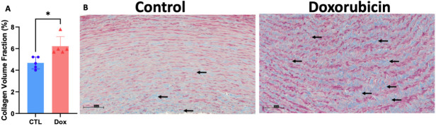
{"title":"Arterial effects of anthracycline: structural and inflammatory assessments in non-human primates and lymphoma patients.","authors":"Stephen Rankin, Caitlin Fountain, Alastair J Gemmell, Daire Quinn, Alasdair Henderson, John McClure, Sandy Small, Balaji Venugopal, Pamela McKay, Piotr J Slomka, David Colville, Mark C Petrie, Giselle C Meléndez, Ninian N Lang","doi":"10.1042/CS20241529","DOIUrl":null,"url":null,"abstract":"<p><p>Anthracyclines, such as doxorubicin, are important anti-cancer therapies but are associated with arterial injury. Histopathological insights have been limited to small animal models, and the role of inflammation in the arterial toxic effects of anthracycline is unclear in humans. Our aims were (1) to evaluate aortic media fibrosis and injury in non-human primates treated with anthracyclines; (2) to assess the effect of anthracycline on aortic inflammation in patients treated for lymphoma. African Green monkeys (AGMs) received doxorubicin (30-60 mg/m2/biweekly intravenously, cumulative dose: 240 mg/m2). Blinded histopathologic analyses of the ascending aorta were performed 15 weeks after the last doxorubicin dose and compared to five age- and gender-matched healthy, untreated AGMs. Analysis of the thoracic aorta of patients with diffuse large B-cell lymphoma (DLBCL), at baseline and after doxorubicin exposure, was performed using 18F-fluorodeoxyglucose (18F-FDG) positron emission tomography/computed tomography (PET/CT) in this observational study by maximal tissue-to-background ratio (TBRmax). In AGMs, doxorubicin exposure was associated with greater aortic fibrosis (collagen deposition: doxorubicin 6.23 ± 0.88% vs. controls 4.67 ± 0.54%; P=0.01) and intracellular vacuolization (doxorubicin 66.3 ± 10.1 vs. controls 11.5 ± 4.2 vacuoles/field, P<0.0001) than untreated controls. In 101 patients with DLBCL, there was no change in aortic TBRmax after anthracycline exposure (TBRmax 1.46 ± 0.16 vs. 1.44 ± 0.14, respectively, P=0.14). Univariate analyses yielded similar results. In a large animal model, anthracycline exposure was associated with aortic fibrosis. In patients with lymphoma, anthracycline exposure was not associated with aortic inflammation. Further research is required to elucidate the mechanisms of anthracycline-related vascular harm.</p>","PeriodicalId":10475,"journal":{"name":"Clinical science","volume":" ","pages":"29-41"},"PeriodicalIF":7.7000,"publicationDate":"2025-01-15","publicationTypes":"Journal Article","fieldsOfStudy":null,"isOpenAccess":false,"openAccessPdf":"https://www.ncbi.nlm.nih.gov/pmc/articles/PMC12203989/pdf/","citationCount":"0","resultStr":null,"platform":"Semanticscholar","paperid":null,"PeriodicalName":"Clinical science","FirstCategoryId":"3","ListUrlMain":"https://doi.org/10.1042/CS20241529","RegionNum":2,"RegionCategory":"医学","ArticlePicture":[],"TitleCN":null,"AbstractTextCN":null,"PMCID":null,"EPubDate":"","PubModel":"","JCR":"Q1","JCRName":"MEDICINE, RESEARCH & EXPERIMENTAL","Score":null,"Total":0}
引用次数: 0
Abstract
Anthracyclines, such as doxorubicin, are important anti-cancer therapies but are associated with arterial injury. Histopathological insights have been limited to small animal models, and the role of inflammation in the arterial toxic effects of anthracycline is unclear in humans. Our aims were (1) to evaluate aortic media fibrosis and injury in non-human primates treated with anthracyclines; (2) to assess the effect of anthracycline on aortic inflammation in patients treated for lymphoma. African Green monkeys (AGMs) received doxorubicin (30-60 mg/m2/biweekly intravenously, cumulative dose: 240 mg/m2). Blinded histopathologic analyses of the ascending aorta were performed 15 weeks after the last doxorubicin dose and compared to five age- and gender-matched healthy, untreated AGMs. Analysis of the thoracic aorta of patients with diffuse large B-cell lymphoma (DLBCL), at baseline and after doxorubicin exposure, was performed using 18F-fluorodeoxyglucose (18F-FDG) positron emission tomography/computed tomography (PET/CT) in this observational study by maximal tissue-to-background ratio (TBRmax). In AGMs, doxorubicin exposure was associated with greater aortic fibrosis (collagen deposition: doxorubicin 6.23 ± 0.88% vs. controls 4.67 ± 0.54%; P=0.01) and intracellular vacuolization (doxorubicin 66.3 ± 10.1 vs. controls 11.5 ± 4.2 vacuoles/field, P<0.0001) than untreated controls. In 101 patients with DLBCL, there was no change in aortic TBRmax after anthracycline exposure (TBRmax 1.46 ± 0.16 vs. 1.44 ± 0.14, respectively, P=0.14). Univariate analyses yielded similar results. In a large animal model, anthracycline exposure was associated with aortic fibrosis. In patients with lymphoma, anthracycline exposure was not associated with aortic inflammation. Further research is required to elucidate the mechanisms of anthracycline-related vascular harm.
期刊介绍:
Translating molecular bioscience and experimental research into medical insights, Clinical Science offers multi-disciplinary coverage and clinical perspectives to advance human health.
Its international Editorial Board is charged with selecting peer-reviewed original papers of the highest scientific merit covering the broad spectrum of biomedical specialities including, although not exclusively:
Cardiovascular system
Cerebrovascular system
Gastrointestinal tract and liver
Genomic medicine
Infection and immunity
Inflammation
Oncology
Metabolism
Endocrinology and nutrition
Nephrology
Circulation
Respiratory system
Vascular biology
Molecular pathology.
 求助内容:
求助内容: 应助结果提醒方式:


