Faris Rustom, Ezekiel Moroze, Pedram Parva, Haluk Ogmen, Arash Yazdanbakhsh
{"title":"Deep learning and transfer learning for brain tumor detection and classification.","authors":"Faris Rustom, Ezekiel Moroze, Pedram Parva, Haluk Ogmen, Arash Yazdanbakhsh","doi":"10.1093/biomethods/bpae080","DOIUrl":null,"url":null,"abstract":"<p><p>Convolutional neural networks (CNNs) are powerful tools that can be trained on image classification tasks and share many structural and functional similarities with biological visual systems and mechanisms of learning. In addition to serving as a model of biological systems, CNNs possess the convenient feature of transfer learning where a network trained on one task may be repurposed for training on another, potentially unrelated, task. In this retrospective study of public domain MRI data, we investigate the ability of neural network models to be trained on brain cancer imaging data while introducing a unique camouflage animal detection transfer learning step as a means of enhancing the networks' tumor detection ability. Training on glioma and normal brain MRI data, post-contrast T1-weighted and T2-weighted, we demonstrate the potential success of this training strategy for improving neural network classification accuracy. Qualitative metrics such as feature space and DeepDreamImage analysis of the internal states of trained models were also employed, which showed improved generalization ability by the models following camouflage animal transfer learning. Image saliency maps further this investigation by allowing us to visualize the most important image regions from a network's perspective while learning. Such methods demonstrate that the networks not only 'look' at the tumor itself when deciding, but also at the impact on the surrounding tissue in terms of compressions and midline shifts. These results suggest an approach to brain tumor MRIs that is comparable to that of trained radiologists while also exhibiting a high sensitivity to subtle structural changes resulting from the presence of a tumor.</p>","PeriodicalId":36528,"journal":{"name":"Biology Methods and Protocols","volume":"9 1","pages":"bpae080"},"PeriodicalIF":1.3000,"publicationDate":"2024-11-19","publicationTypes":"Journal Article","fieldsOfStudy":null,"isOpenAccess":false,"openAccessPdf":"https://www.ncbi.nlm.nih.gov/pmc/articles/PMC11631523/pdf/","citationCount":"0","resultStr":null,"platform":"Semanticscholar","paperid":null,"PeriodicalName":"Biology Methods and Protocols","FirstCategoryId":"1085","ListUrlMain":"https://doi.org/10.1093/biomethods/bpae080","RegionNum":0,"RegionCategory":null,"ArticlePicture":[],"TitleCN":null,"AbstractTextCN":null,"PMCID":null,"EPubDate":"2024/1/1 0:00:00","PubModel":"eCollection","JCR":"Q3","JCRName":"BIOCHEMICAL RESEARCH METHODS","Score":null,"Total":0}
引用次数: 0
Abstract
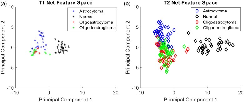
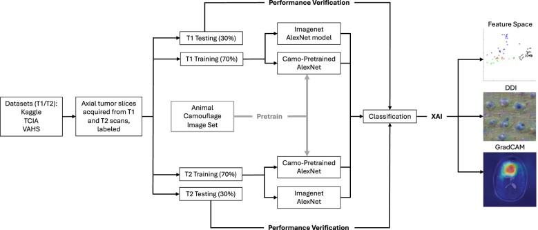
Convolutional neural networks (CNNs) are powerful tools that can be trained on image classification tasks and share many structural and functional similarities with biological visual systems and mechanisms of learning. In addition to serving as a model of biological systems, CNNs possess the convenient feature of transfer learning where a network trained on one task may be repurposed for training on another, potentially unrelated, task. In this retrospective study of public domain MRI data, we investigate the ability of neural network models to be trained on brain cancer imaging data while introducing a unique camouflage animal detection transfer learning step as a means of enhancing the networks' tumor detection ability. Training on glioma and normal brain MRI data, post-contrast T1-weighted and T2-weighted, we demonstrate the potential success of this training strategy for improving neural network classification accuracy. Qualitative metrics such as feature space and DeepDreamImage analysis of the internal states of trained models were also employed, which showed improved generalization ability by the models following camouflage animal transfer learning. Image saliency maps further this investigation by allowing us to visualize the most important image regions from a network's perspective while learning. Such methods demonstrate that the networks not only 'look' at the tumor itself when deciding, but also at the impact on the surrounding tissue in terms of compressions and midline shifts. These results suggest an approach to brain tumor MRIs that is comparable to that of trained radiologists while also exhibiting a high sensitivity to subtle structural changes resulting from the presence of a tumor.

 求助内容:
求助内容: 应助结果提醒方式:


