{"title":"Lung Point in a Case of Bronchoscopy Lung Volume Reduction: Consider Its Mimics Before Inserting the Tube.","authors":"Mohannad Wazirali, Paul M Shaniuk","doi":"10.24908/pocus.v9i2.17551","DOIUrl":null,"url":null,"abstract":"<p><p>Point of Care Ultrasound (POCUS) is used to evaluate many clinical scenarios. Chest POCUS has been integrated as a part of a clinical protocol to assess patients with lung pathology 1. The ability to detect pneumothorax using chest POCUS has been shown to be superior to chest radiography, with specificity reported to be as high as 100% when a lung point sign is identified. In addition to improved diagnostic accuracy, chest POCUS has the added benefits of ease of access and absence of ionizing radiation. Here we describe a case where a patient with a high pre-test probability for pneumothorax had a detected lung point sign, but pneumothorax was ruled out via Computed Tomography (CT). This case highlights the importance of considering the mimics of the lung point sign. This case also shows a unique and interesting finding related to pleural movement restriction post-Bronchoscopic lung volume reduction (BLVR).</p>","PeriodicalId":74470,"journal":{"name":"POCUS journal","volume":"9 2","pages":"46-49"},"PeriodicalIF":0.0000,"publicationDate":"2024-11-15","publicationTypes":"Journal Article","fieldsOfStudy":null,"isOpenAccess":false,"openAccessPdf":"https://www.ncbi.nlm.nih.gov/pmc/articles/PMC11614493/pdf/","citationCount":"0","resultStr":null,"platform":"Semanticscholar","paperid":null,"PeriodicalName":"POCUS journal","FirstCategoryId":"1085","ListUrlMain":"https://doi.org/10.24908/pocus.v9i2.17551","RegionNum":0,"RegionCategory":null,"ArticlePicture":[],"TitleCN":null,"AbstractTextCN":null,"PMCID":null,"EPubDate":"2024/1/1 0:00:00","PubModel":"eCollection","JCR":"","JCRName":"","Score":null,"Total":0}
引用次数: 0
Abstract
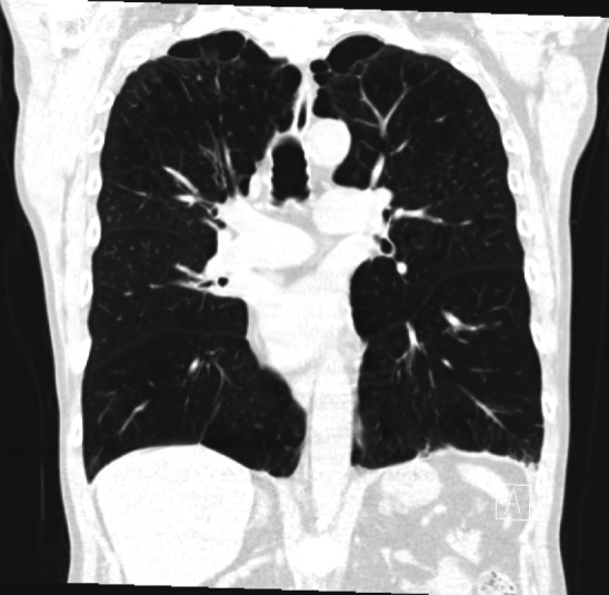
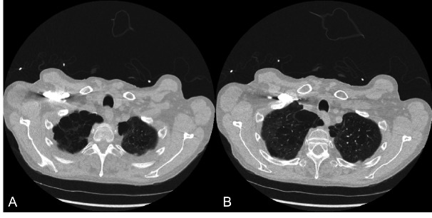
Point of Care Ultrasound (POCUS) is used to evaluate many clinical scenarios. Chest POCUS has been integrated as a part of a clinical protocol to assess patients with lung pathology 1. The ability to detect pneumothorax using chest POCUS has been shown to be superior to chest radiography, with specificity reported to be as high as 100% when a lung point sign is identified. In addition to improved diagnostic accuracy, chest POCUS has the added benefits of ease of access and absence of ionizing radiation. Here we describe a case where a patient with a high pre-test probability for pneumothorax had a detected lung point sign, but pneumothorax was ruled out via Computed Tomography (CT). This case highlights the importance of considering the mimics of the lung point sign. This case also shows a unique and interesting finding related to pleural movement restriction post-Bronchoscopic lung volume reduction (BLVR).


 求助内容:
求助内容: 应助结果提醒方式:


