The perspectives of neurologists on positron emission tomography utility in multiple sclerosis: A qualitative study
IF 2.9
3区 医学
Q2 CLINICAL NEUROLOGY
引用次数: 0
Abstract
Background
Magnetic resonance imaging (MRI) is the gold standard for imaging disease activity in multiple sclerosis (MS) patients. However, recent studies indicate that positron emission tomography (PET) may provide added value in visualizing MS disease in the future.
Objective
This study aims to investigate the barriers to implementing PET for MS patients and its potential added value in the context of MS.
Methods
11 semi-structured in-depth interviews with neurologists specialized in MS were conducted. The neurologists were selectively recruited from six medical centers in Belgium and the Netherlands. Inductive thematic analysis was used to analyze the data.
Results
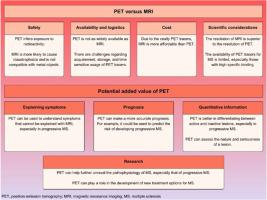
The interviews revealed several hurdles that play a role in using PET for MS, including financial and scientific considerations. Potential clinical applications of PET were also identified, such as understanding unexplained symptoms, making a more accurate prognosis, evaluating the nature and seriousness of a lesion, and assessing disease activity. In addition, research applications were highlighted, including unraveling the pathophysiology of MS and developing new treatment options for MS.
Conclusion
Using PET is advancing our understanding of MS and can accelerate the development of novel therapies to combat its progression. However, its integration into routine clinical practice for MS remains a future prospect, contingent upon further technological advancements and supportive healthcare frameworks.

神经科医生对正电子发射断层扫描在多发性硬化症中的应用的看法:定性研究
背景磁共振成像(MRI)是多发性硬化症(MS)患者疾病活动成像的黄金标准。然而,最近的研究表明,正电子发射断层扫描(PET)可能会在未来为多发性硬化症疾病的可视化提供附加值。本研究旨在调查多发性硬化症患者实施 PET 的障碍及其在多发性硬化症方面的潜在附加值。这些神经科医生是从比利时和荷兰的六个医疗中心有选择地招募的。结果访谈揭示了将 PET 用于多发性硬化症的几个障碍,包括财务和科学方面的考虑。还发现了 PET 的潜在临床应用,如了解不明原因的症状、做出更准确的预后、评估病变的性质和严重程度以及评估疾病活动。此外,研究应用也得到了强调,包括揭示多发性硬化症的病理生理学和开发治疗多发性硬化症的新方案。然而,将 PET 纳入多发性硬化症的常规临床实践仍是未来的一个前景,这取决于进一步的技术进步和支持性的医疗保健框架。
本文章由计算机程序翻译,如有差异,请以英文原文为准。
求助全文
约1分钟内获得全文
求助全文
来源期刊

Multiple sclerosis and related disorders
CLINICAL NEUROLOGY-
CiteScore
5.80
自引率
20.00%
发文量
814
审稿时长
66 days
期刊介绍:
Multiple Sclerosis is an area of ever expanding research and escalating publications. Multiple Sclerosis and Related Disorders is a wide ranging international journal supported by key researchers from all neuroscience domains that focus on MS and associated disease of the central nervous system. The primary aim of this new journal is the rapid publication of high quality original research in the field. Important secondary aims will be timely updates and editorials on important scientific and clinical care advances, controversies in the field, and invited opinion articles from current thought leaders on topical issues. One section of the journal will focus on teaching, written to enhance the practice of community and academic neurologists involved in the care of MS patients. Summaries of key articles written for a lay audience will be provided as an on-line resource.
A team of four chief editors is supported by leading section editors who will commission and appraise original and review articles concerning: clinical neurology, neuroimaging, neuropathology, neuroepidemiology, therapeutics, genetics / transcriptomics, experimental models, neuroimmunology, biomarkers, neuropsychology, neurorehabilitation, measurement scales, teaching, neuroethics and lay communication.
 求助内容:
求助内容: 应助结果提醒方式:


